Download
1 / 89
991 likes | 1.49k Views
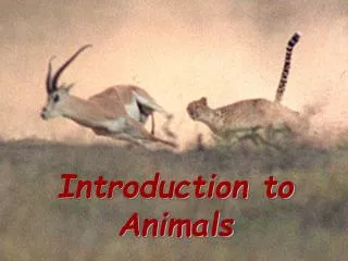
SUPPORT & MOVEMENT IN ANIMALS. Why is locomotion important to animals? To escape unfavourable conditions , e.g . predators, To find food ; To seek mates ; To disperse to new habitats; To seek favourable envi ronments ; e.g. shelter. Locomotion in Unicellular Organisms.

E N D
SUPPORT & MOVEMENT IN ANIMALS Why is locomotion important to animals? • To escape unfavourable conditions, e.g. predators, • To find food; • To seek mates; • To disperseto new habitats; • To seek favourable environments; e.g. shelter
Locomotion in Unicellular Organisms Involves using; • Pseudopodia e.g. amoeba. • flagellume.g. Trypanosoma spp. • ciliae.g. Paramecium spp.
Locomotion in Multicellular organisms Requires; • Muscles, a contractile tissue, which provide a source of power • Skeleton, on which muscles can act to bring movement
Types of Skeleton • Hydrostatic (hydraulic), • Exoskeleton, • Endoskeleton,
Hydrostatic (hydraulic) • Mechanical support is provided by an internal fluid-filled system. • E.g. most invertebrates like earthworm, leeches, caterpillars and maggots.
Exoskeleton • E.g. arthropods – made of chitin, • Exoskeleton forms a hard casing enclosing the softer tissues of the arthropod body, • Jointed limbs movements are due to antagonistic muscles, • The Muscles are attached internally,
Endoskeleton E.g. chordates made up of; • bones, • cartilage tissue,
General Functions of the Endoskeleton The Endoskeleton has nine main functions: • Provide shape and support, • Provide Attachment, • Provide a frame work for Movement, • Provide Protection, • Site of Blood cell production, • Provide Storage, • Involved in pH buffering, • Involved in Detoxification, • Involved in Sound transduction,
The Mammalian Skeleton Major components include: • the axial skeleton, • the appendicular skeleton,
axial skeleton The axial skeleton has five areas; • Skull, • Ossicles bones, • Hyoid bone in the throat, • Vertebral column, • Chest,
Appendicular Skeleton The appendicular skeleton consists of; • the limb bones, • the pelvic girdle, • pectoralgirdle,
Skull The Skull: • Consists of cranium, facial bones and two jaws, • At the base of the cranium, are occipital condyles which articulate with the first vertebral bone, atlas, Functions of the Skull; • Mechanical protection of brain & sensory organs. • Upper & lower jaws used for chewing food.
Front view & Lateral view of Human skull Cranium bones include; Nasal bones, Frontal bones, Parietal bones, Temporal bones, Zygomatic arches, Occipital bones,
THE VERTEBRAL COLUMN Backbone consists of Vertebrae (Singular- vertebra) grouped into; Cervical vertebrae Thoracic vertebrae, Lumbar vertebrae, Sacral vertebrae, Caudal vertebrae (coccyx),
A TYPICAL VERTEBRA A main parts of typical vertebra; neural canal- passage of spinal cord, Neural arch- surrounds neural canal, Neural spine- projects upwards/dorsally, Centrum (plural-centra)- ventrally located and fits into intervertebral discs on both sides Transverse processes- on either side of neural arch, Zygapophyses(singular-zygapophysis)- articulation smooth facets with adjacent vertebra,
Cervical Vertebrae Comprising of ; • 1st Cervical vertebra (Atlas vertebra), • 2nd Cervical vertebra (Axis vertebra), • 3rd -7th cervical vertebrae,
Distinguishing features of cervical vertebrae All 7 cervical vertebrae have; • vertebrarterial canals, • transverse processes flattened out to form cervical ribs, • large neural cavity, • smallcentrum,
Atlas vertebra dorsal view Features of the Atlas vertebra; Very large Neural canal, Prominent cervical ribs (transverse processes), Large hollow facets (articulate with occipital condyles) Reduced centrum, Reduced neural spine, Large Postzygapophyses to articulate with prezygapophyses of axis,
Atlas anterior view Showing the articulation surface with the occipital condyles of the skull
Axis vertebra lateral view Features of Axis vertebra; Large centrum forming Odontoid process, large neural spine, Flat cervical ribs, postzygapophyses
3rd – 7th Cervical vertebra anterior view All 7 cervical vertebrae have; vertebrarterial canals, transverse processes flattened out to form cervical ribs, large neural cavity, small centrum,
Adaptations of cervical vertebrae • broad neural arch for protection of the spinal cord. • forked and short transverse processes for the attachment of neck muscles. • Atlas has broad surfaces for articulation with the occipital condyles of the skull to permit nodding movement of the skull. • vertebrarterial canals for passage of neck blood vessels and nerves.
continued • Axis has odontoid process; a projection of the centrum to permit rotator movement of the skull. • The odontoid process acts as a pivot; for the atlas and skull. • short neural spine for attachment of neck muscles. • wide neural canal for passage of the enlarged spinal cord.
Thoracic Vertebrae (lateral view) Distinguishing features of a thoracic vertebra; Long neural spine projecting upwards & backwards, Short transverse processes, Tubercular facet (on ventral side of transverse processes), Capitular demi-facet, Other features; Large centrum, Large neural canal, Prezygapophyses, Postzygapophyses, Neural canal,
Adaptations of thoracic vertebrae • neural arch for protection of the spinal cord. • centrum for attachment of the transverse processes. • pre and post zygapophyses facets for articulation with those of the next vertebrae.
Continued • tubercular and capitular facets for articulation with the tuberculum and capitulum of the rib, • reduced transverse processes for attachment of muscles, • long neural spine for attachment of the back muscles,
Lumbar Vertebrae (Anterior view) Distinguishing features of a lumbar vertebra; Broad neural spine pointing upwards & forward, Large, thick centrum,(supporting the weight of the animal), Large transverse processes, (abdominal muscle attachment), Metapophyses, (abdominal muscle attachment), Anapophyses, (abdominal muscle attachment), Hypapophyses, (abdominal muscle attachment),
Adaptation of lumbar • broad neural spine for attachment of powerful back and abdominal muscles. • long and well developed transverse processes for attachment of muscles that maintain posture and flexes the spine. • metapophyses projections provide additional surface for muscle attachment.
continued • hypapophyes projections provide additional surface for muscle attachment. • Thick and compact centrum for support. • pre and post zygapophyses for articulation between the vertebrae
SACRAL VERTEBRAE (Ventral view) Distinguishing features of Sacral vertebrae; sacral vertebrae are fused to form,sacrum, 1st Sacral vertebrae transverse processes are large & fused with the pelvic girdle, Numerous foramen (canals), Reduced metapophyses, Large centrum, Narrow neural canal, neural spine reduce posteriorly,
Sacral vertebrae Sacrum dorsal View Sacrum lateral view
Adaptations of the Sacral Vertebrae • Numerous canals for passage of blood vessels and nerves, • Sacral vertebrae are fused to provide strength and firmness,
continued • The 1stsacral vertebra has well developed transverse processes, which are fused to the pelvic girdle to provide support and mechanical protection to lower abdomen organs. • The 1st sacral vertebra has Large neural spine & transverse processes provide attachment to lower back & thigh muscles,
Coccyx (caudal) vertebrae Consists of four coccygeal vertebrae, • Neural spine, transverse processes & neural canal reduced,
Ribs Twelve ribs; Seven true ribs, Three False ribs, Two floating, The rib has two parts; Vertebral part; bearing tuberculum & capitulum,, Sternal part;
sternum Consists of three sections; • Manubrium articulates with 1st two pairs of ribs, • Body articulates to ribs 3rd-7th , • Xiphoid cartilage supports abdominal muscles,