Download
1 / 48
580 likes | 2.28k Views
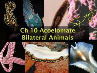
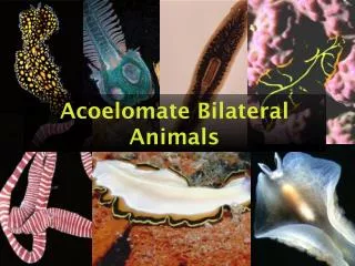
Acoelomate Bilateral Animals. Acoelomate Bilateral Animals. Consist of phyla: Phylum Platyhelminthes Phylum Nemertea And others. Reproductive and osmoregulatory systems. Acoelomate Bilateral Animals. Simplest organisms to have bilateral symmetry Triploblastic Lack a coelom

E N D
Acoelomate Bilateral Animals • Consist of phyla: • Phylum Platyhelminthes • Phylum Nemertea • And others
Reproductive and osmoregulatory systems Acoelomate Bilateral Animals • Simplest organisms to have bilateral symmetry • Triploblastic • Lack a coelom • Organ-system level of organization • Cephalization • Elongated, without appendages
Bilateral Symmetry • Divided along sagittal plane into two mirror images • sagittal= divides bilateral organisms into right and left halves
Review • Anterior= head end • Posterior= tail end • Dorsal= back side • Ventral= belly side
Bilateral animals • Bilateral symmetry = important evolutionary advancement • Important for active, directed movement • Anterior, posterior ends • One side of body kept up (dorsal) vs. down (ventral)
Directed movement evolved with anterior sense organscephalization Cephalization • specialization of sense organs in head end of animals
Acoelomate Phylum Platyhelminths Or not shown here Nemerterean • Acoelomates lack a true body cavity • Solid body • no cavity b/w the digestive tract and outer body wall This is a round worm Different Phylum
Acoelomates are triploblastic • Triploblastic (3 germ layers) • Germ layer= layers in embryo that form the various tissues and organs of an animal body
3 germ layers • Ectoderm • Outermost germ layer • Gives rise to outer covering of animal ie. epidermis • Endoderm • Innermost germ layer • Gives rise to inner lining of gut tract
Mesoderm • Middle germ layer • b/w ectoderm and endoderm • Gives rise to various tissues/organs (ie. muscles)
Acoelomate animals have an organ-system level of organization
Digestive tract and nervous system Acoelomate animals have an organ-system level of organization • Organ-system • Different organs operate together (ie. excretory system, nervous system) • mesodermal tissue gives rise to parenchyma
Polyclad • From Red Sea http://www.rzuser.uni-heidelberg.de/~bu6/
Phylum Platyhelminthes Flatworms Free living Parasitic
From Atlantic ocean http://www.rzuser.uni-heidelberg.de/~bu6/
Phylum Platyhelminthes • Flattened dorsoventrally • flatworms • 34,000 species • Gastrovascular cavity (if present) has only one opening (mouth = anus) • Mostly monoecious
Phylum Platyhelminthes • First phylum that has an Organ systems present • derived mesodermally (parenchyma): • Muscular system • Digestive system (incomplete; gastrovascular type) (absent in some) • Nervous system • Excretory system (absent in some) • Reproductive system
Rely on diffusion Phylum Platyhelminthes • Organ systems absent: • Circulatory • Respiratory
Hymenolepsis- rat tapeworm Phylum Platyhelminthes (cont’d) • Divided into 4 classes: • Class Turbellaria (mostly free-living flatworms) • Class Trematoda (parasitic flukes) • Class Monogenea (ectoparasitic flukes) • Class Cestoda (tapeworms)
Class Turbellaria • Mostly free-living flatworms • Marine (mostly) or freshwater bottom-dwellers • Predators and scavengers • First group of bilateral symmetrical animals Planarian genus Dugesia
Digestion is completed within the cells lining the gastro- vascular cavity, which has three branches, each with fine subbranches that pro- vide an extensive surface area. Pharynx. The mouth is at the tip of a muscular pharynx that extends from the animal’s ventral side. Digestive juices are spilled onto prey, and the pharynx sucks small pieces of food into the gastrovascular cavity, where digestion continues. Undigested wastes are egested through the mouth. Gastrovascular cavity Eyespots Ganglia. Located at the anterior end of the worm, near the main sources of sensory input, is a pair of ganglia, dense clusters of nerve cells. Ventral nerve cords. From the ganglia, a pair of ventral nerve cords runs the length of the body. Figure 33.10 • The best-known turbellarians, commonly called planarians • Have light-sensitive eyespots and centralized nerve nets
Class Turbellaria (cont’d) • Move by muscles, ciliated epidermis /gastrovascular cavity
Class Turbellaria (cont’d) • Freshwater turbellarians adapted osmoregulatory structures • Protonephridia • protos= first • nephros= kidney • network of fine tubules running down sides of organism
Class Turbellaria (cont’d) • Flame cells= branch from tubules • Ciliary projections drive fluid down tubule • Tubules open to outside= nephridiopore
Class Turbellaria (cont’d) • nervous system with nerve ganglion • ganglion- aggregation of nervous tissue • Cephalization- cerebral ganglion= primitive brain
Class Turbellaria (cont’d) • Ocelli= light-sensitive eyespots
Reproductive and osmoregulatory systems Turbellarian Reproduction • Asexual (fission) • transverse • Sexual • Monoecious (mostly) • Cross-fertilization
All parasitic • lack cilia • Have unusual body covering: tegument Other 3 classes: • Class Trematoda • Class Monogenea • Class Cestoda • Outer zone of tegument (glycocalyx) • consists of proteins and carbohydrates • aids in transport of nutrients, waste, gases • Protection against host defenses
Class Trematoda • Parasitic flukes • Endoparasites • Hooks, suckers, increased reproductive capacity
1mm-6cm long • Complex life cycle: • Definitive host (primary/final host) • where parasite matures and reproduces (sexually) (eggs released) • vertebrate
Intermediate host • Mollusc (ie. snail) • Hosts in which larval stages develop and undergo asexual reproduction • Results in an increase in the number of the individuals
Mature flukes live in the blood vessels of the human intestine. A female fluke fits into a groove running the length of the larger male’s body, as shown in the light micrograph at right. 1 Male Female 1 mm 5 These larvae penetrate the skin and blood vessels of humans working in irrigated fields contaminated with infected human feces. Blood flukes reproduce sexually in the human host. The fertilized eggs exit the host in feces. 2 3 The eggs develop in water into ciliated larvae. These larvae infect snails, the intermediate hosts. Asexual reproduction within a snail results in another type of motile larva, which escapes from the snail host. 4 Snail host Figure 33.11 • Trematodes that parasitize humans • Spend part of their lives in snail hosts
Chinese Liver Fluke • Infects 30 million people in eastern Asia • Lives in ducts of liver • Eats epithelial tissue, blood • Definitive host: • Humans, dogs, cats • 2 intermediate hosts: • snail • fish
Class Monogenea • Parasitic flukes • Mostly ectoparasites • Single host, mostly fish
Hymenolepsis- rat tapeworm Class Cestoda • Tapeworms • Endoparasites • Vertebrate host • Live in digestive tract • 1 mm- 25m long (EWWWW!!)
Proglottids with reproductive structures 200 µm Hooks Scolex Sucker Figure 33.12 Tapeworm • Tapeworms • Are also parasitic and lack a digestive system
Hymenolepsis- rat tapeworm Class Cestoda • Highly specialized • Lack mouth, digestive tract • Absorb nutrients across body wall • Hooks and suckers • “head”= scolex
Adult tapeworms consist of long series of repeating units= proglottids • Chain of proglottids= strobila
Tapeworms are monoecious (mostly) • Mostly cross-fertilization • No specialized sense organs scolex
Cestodes depend on host digestion • Small molecules in host intestine, liver
Beef Tapeworm • Definitive host= human • Intermediate host= cattle
Phylum Nemertea- ribbon worm • Triplobastic, acoelamate • bilateral symmetry • Unsegmented • Ciliated epidermis • Closed circulatory • usually <20cm • Marine mud, sand • Elongate, flattened worms
Phylum Nemertea (cont’d) • Unlike the platyhelminthes, Complete digestive tract, with anus • One-way • More efficient; allows larger growth
Phylum Nemertea (cont’d) • Cerebral ganglion, longitudinal nerve cords • Long proboscis used in carnivorous species • Two lateral blood vessels yet no heart • Dioecious • “two” “house” • Male and female organs in separate individuals