Download
1 / 90
1.25k likes | 2.36k Views
Protection, Support, and Movement. Chapter 39. KEY CONCEPTS. Many structures and processes have evolved in animals for protection, support, and movement. Learning Objective 1. Compare the functions of the external epithelium of invertebrates and vertebrates. Epithelial Tissue.

E N D
Protection, Support, and Movement Chapter 39
KEY CONCEPTS • Many structures and processes have evolved in animals for protection, support, and movement
Learning Objective 1 • Compare the functions of the external epithelium of invertebrates and vertebrates
Epithelial Tissue • In both invertebrates and vertebrates • protects underlying tissues • specialized sensory or respiratory functions • Outer epithelium specialized to secrete • lubricants or adhesives • odorous or poisonous substances
Epithelial Tissuein Invertebrates • Cuticle • protective shell secreted by outer epithelium
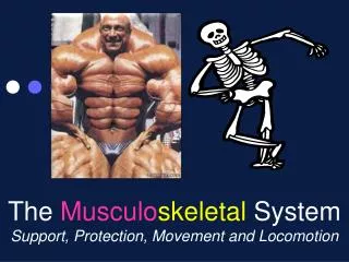
Integumentary System of Vertebrates • Skin and structures that develop from it • Mammalian skin includes • hair, claws or nails, sweat glands, oil glands, sensory receptors
Learning Objective 2 • Relate the structure of vertebrate skin to its functions
Feathers and Hair • Feathers of birds and hair of mammals • form insulating layer • helps maintain constant body temperature
Epidermis 1 • Protects body from outer environment • Stratum corneum • most superficial layer • consists of dead cells filled with keratin • Keratin • insoluble protein • gives mechanical strength to skin • reduces water loss
Epidermis2 • Stratum basale • cells divide, are pushed up to skin surface • cells mature, flatten, produce keratin • eventually die and slough off
Dermis • Consists of dense, fibrous connective tissue • Rests on layer of subcutaneous tissue • composed largely of insulating fat
Openings of sweat glands Nerve endings Capillary Stratum corneum Melanocyte (pigment cell) Epidermis Stratum basale Hair erector muscle Dermis Hair shaft Sensory receptor (Pacinian corpuscle) Subcutaneous tissue Artery Hair follicle Sweat gland Vein Sebaceous gland Fig. 39-1, p. 829
KEY CONCEPTS • Epithelial coverings protect underlying tissues and may be specialized for sensory, respiratory, or other functions
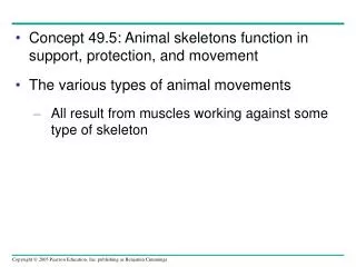
Learning Objective 3 • Compare the structure and functions of different types of skeletal systems, including the hydrostatic skeleton, exoskeleton, and endoskeleton
TheSkeletal System • Supports and protects the body • Transmits mechanical forces generated by muscles
Hydrostatic Skeleton • Fluid in closed body compartment • transmits forces generated by contractile cells or muscle • Found in soft-bodied invertebrates • cnidarians, flatworms, annelids
Longitudinal contractile fibers of epidermal layer Circular contractile fibers of gastrodermis Contraction of circular contractile fibers elongates the body. (b) Contraction of longitudinal fibers shortens the body. Fig. 39-2, p. 830
Longitudinal contractile fibers of epidermal layer Circular contractile fibers of gastrodermis Contraction of circular contractile fibers elongates the body. (b) Contraction of longitudinal fibers shortens the body. Stepped Art Fig. 39-2, p. 830
Exoskeletons • Nonliving skeleton • characteristic of mollusks and arthropods • doesn’t grow, arthropods must molt periodically • Arthropod skeleton • composed partly of chitin • jointed for flexibility • adapted for many lifestyles
Endoskeletons • Consist of living tissue • can grow • Found in echinoderms and chordates
Learning Objective 4 • Describe the main divisions of the vertebrate skeleton and the bones that make up each division
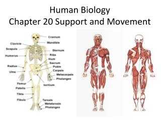
The Vertebrate Skeleton 1 • Axial skeleton • skull • vertebral column • rib cage • sternum
The Vertebrate Skeleton 2 • Appendicular skeleton • limbs • pectoral girdle • pelvic girdle
Skull Sternum Rib cage Vertebrae Axial skeleton (brown) Fig. 39-5a, p. 832
Clavicle Scapula Humerus Pelvic girdle Radius Ulna Carpals Metacarpals Phalanges Femur Patella Fibula Tibia Tarsals Metatarsals Phalanges Appendicular skeleton (brown) Fig. 39-5b, p. 832
KEY CONCEPTS • Skeletal systems, whether they are hydrostatic skeletons, exoskeletons, or endoskeletons, support and protect the body and transmit mechanical forces important in movement
Learning Objective 5 • Describe the structure of a typical long bone • Differentiate between endochondral and intramembranous bone development
A Long Bone • Consists of • a thin outer shell of compact bone surrounding inner spongy bone • a central cavity that contains bone marrow
Articular surface covered with cartilage Epiphysis Red marrow in spongy bone Metaphysis Periosteum Yellow marrow Blood supply Diaphysis Compact bone Articular cartilage Epiphysis Fig. 39-6, p. 833
Bone Development • Long bones • develop from cartilage templates during endochondral bone development • Other bones (such as flat bones of skull) • develop from noncartilage connective tissue model by intramembranous bone development
Bone Cells • Osteoblasts • cells that produce bone • Osteoclasts • cells that break down bone • Osteoblasts and osteoclasts work together to shape and remodel bone
Learn more about the human skeletal system and a typical long bone by clicking on the figures in ThomsonNOW.
Learning Objective 6 • Compare the main types of vertebrate joints
Joints • Junctions of two or more bones • Ligaments • connective tissue bands • connect bones • limit movement in joint
Types of Joints • Immovable joints • sutures of the skull • Slightly movable joints • joints between vertebrae • Freely movable joint • enclosed by joint capsule lined with membrane that secretes synovial fluid
Learning Objective 7 • Relate the structure and function of insect flight muscles
Insect Flight Muscles • Large numbers of mitochondria and tracheae (air tubes) • support high metabolic rate required for flight
Learning Objective 8 • Describe the structure of skeletal muscles and their antagonistic actions
Muscular Systems • In vertebrates and most invertebrates • muscle tissue contracts (shortens) • moves body parts by pulling on them • Three types of muscle • skeletal • smooth • cardiac muscle
Facial muscles Muscles that flex fingers Sternocleido- mastoid Platysma Trapezius Clavicle Latissimus dorsi Deltoid Pectoralis major Rectus abdominis Biceps brachii Linea alba Brachialis External oblique Gluteus medius Wrist and finger flexors Gracilis Sartorius Triceps brachii Quadriceps femoris Patella Gastrocnemius Tibialis anterior Tibia Soleus Fig. 39-8a, p. 835
Sternocleidomastoid Biceps brachii Trapezius Deltoid Brachialis Triceps brachii Latissimus dorsi External oblique Brachioradialis Muscles that flex fingers Gluteus maximus Gracilis Semitendinosus Hamstring muscles Biceps femoris Semi-membranosus Gastrocnemius Soleus Achilles tendon Calcaneus Fig. 39-8b, p. 835
Vertebrate Skeletal Muscles • Pull on tendons • connective tissue, attaches muscles to bones • Muscle contraction • pulls bone toward or away from the bone with which it articulates
Muscle Actions • Skeletal muscles act antagonistically to one another • Agonist • muscle that produces a particular action • Antagonist • produces the opposite movement