
Practical Guide to Gram Staining: Reagents, Procedure, and Applications
E N D
Presentation Transcript
PRACTICALGRAM STAINING Assist Prof Dr. Syed YousafKazmi
OBJECTIVES • Describe reagents used in Gram stain & purpose of these reagents • Color expected of Gram Pos & Gram Neg after performing the procedure • Explain reason of differential stain by Gram Pos & Gram Neg • Describe cell wall structure of Gram Pos & Neg • Explain Gram variable org
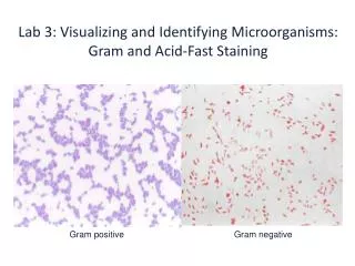
REAGENTS USED IN GRAM STAIN • Gram Crystal Violet 0.5% • Gram Iodine • Potassium Iodide 2% • Resublimed Iodine 1% • Gram Decolorizer • Methanol 80% • Acetone 20% • Gram Safranine1% Gram negative Gram positive
REAGENTS USED IN GRAM STAIN • CRYSTAL VIOLET • Primary stain • Violet colored, stains all micro-org • GRAM IODINE • Mordant • Forms Crystal violet iodine complexes • DECOLORIZER • Acetone + Methanol • Removes Crystal violet iodine complex from thin peptidoglycan layers • Dissolves outer layer of Gram negative org
REAGENTS USED IN GRAM STAIN • GRAM SAFRANINE • Counter stain • Red colored • Stains thin walled Gram neg org • Pus cells cytoplasm & lobes of nuclie also stain red
The Gram Stain Procedure Step 1 - Prepare aSmear Suspend some of the material to be stained in a drop of water on a microscope slide, spread the drop to about the size of a nickel. Allow to air dry. Heat fix by gently warming Watch what happens to the “Bacteria” at each step “Bacteria”
The Gram Stain Procedure Step 2 - Apply the Primary Stain Flood the Smear with Crystal Violet Allow to stand for 1 min Rinse with water to remove excess stain
The Gram Stain Procedure Step 3 - Apply the Mordant Flood the Smear with Iodine solution Allow to stand 2 min
The Gram Stain Procedure Step 4 - Rinse Rinse with water to remove excess Iodine
The Gram Stain Procedure Step 5 - Decolorize Drip Decolorizer (80% Methanol +20% Acetone) across the slide about 5 sec The effluent should appear pale or clear
The Gram Stain Procedure Step 6 - Rinse Rinse with water to remove excess alcohol
The Gram Stain Procedure Step 7 - Counterstain Flood the slide with Safranin solution Let stand for 2 minutes
The Gram Stain Step 8 - Rinse,Dry and Observe Rinse with water to remove excess stain Blot dry Observe under Oil Immersion Gram-Positive Gram-Negative
GRAM VARIABLE • Gram variability • Old cultures • Decolorize improperly • Dead and alive bacteria • Autolytic organisms e.g. Streptococcus pneumoniae