Download
1 / 20
310 likes | 1.24k Views
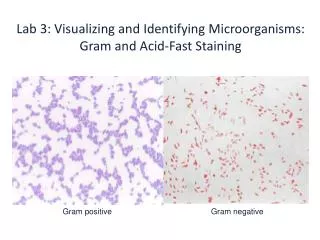
Lab 3: Visualizing and Identifying Microorganisms: Gram and Acid-Fast Staining. Gram positive. Gram negative. Why do we need to stain microorganisms?. Bacterial Smears. Before bacteria can be stained a smear must be made and fixed.

E N D
Lab 3: Visualizing and Identifying Microorganisms: Gram and Acid-Fast Staining Gram positive Gram negative
Bacterial Smears • Before bacteria can be stained a smear must be made and fixed. • A bacterial smear is made by spreading bacteria on a clean slide and allowing it to air dry or place on electric slide warmer. • The slide is passed over the incinerator to heat fix the bacteria.
Preparing a Bacterial Smear • Get clean microscope slide (handle carefully, no fingerprints!) • Vortex (mix) the broth culture until it is cloudy (or add 1-2 drops of water to the slide if the starter culture a solid) • Using a loop, spread a thin film of bacteria (smear) on the slide and let it air dry • Gently heat the slide by placing it near the front of the incinerator or on the electric slide warmer to fix the bacteria onto the slide
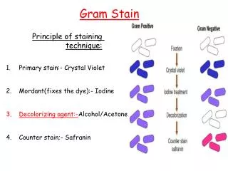
Part 1: The Gram Stain • The Gram Stain is a differential stain used to classify Gram positive and negative bacteria. • Developed by Hans Christian Gram in 1884 when he was studying bacteria from different respiratory diseases. • The single most important technique in microbiology.
Reagents for Gram Staining • Crystal violet (CV): primary stain; all bacteria stain blue. • Grams Iodine (I): not a stain; it forms a crystal violet—iodine complex inside the cell wall. • Decolorizer: ethyl alcohol, used to remove the primary stain. It removes the CV-I complex from cells without teichoic acid. • Safranin: a counter stain; it stains the decolorized cells a red color. What’s the difference in a Gram positive vs. gram negative cell? *Gram positive bacteria have many layers of peptidoglycan in their cell wall making it easier to hold onto the CV-I complex. *Gram negative bacteria have one layer of peptidoglycan. Thus the CV-I complex is not retained in the cell.
The Gram Stain The uptake and retaining of the dye depends on the cell wall.
Gram Stain Lab Procedure The detailed procedure can be found on pages 34-35
Part 2: Acid-Fast Staining • Acid fast is a differential stain that detects acid-fast and non-acid fast organisms. • Acid fast bacteria have a waxy cell wall (mycolic acid) that makes them difficult to stain with traditional methods. Acid-fast positive (red) Acid-fast negative (blue)
Part 2: Acid-Fast Staining Reagents used in Acid-fast staining: • Carbol fuchsin: fuchsin is the red dye and carbol is the acid (carbolic acid) • Acid alcohol: decolorizer • Methylene blue: a blue counter stain Acid-fast positive (red) Acid-fast negative (blue)
Acid-Fast Stain (Kinyoun) Procedure The detailed procedure can be found on pages 41-42
Assignments for this week Lab Reports • Gram stain: all questions • AF stain: drawings and questions 2-4