Staining Methods
General Pathology – Seminary 1b. Staining Methods. Jaroslava Dušková Inst. Pathol. ,1st Med. Faculty, Charles Univ., Prague http://www1.lf1.cuni.cz/~jdusk/. Tissue Samples. Fixation & Embedding FFPE Sections Staining. Cytology Samples. Smears , cytocentrifugation


Staining Methods
E N D
Presentation Transcript
General Pathology – Seminary 1b Staining Methods Jaroslava Dušková Inst. Pathol. ,1st Med. Faculty, Charles Univ., Prague http://www1.lf1.cuni.cz/~jdusk/
Tissue Samples Fixation & Embedding FFPE Sections Staining
Cytology Samples Smears , cytocentrifugation Fixation (formalin, glutaraldehyde, air drying, freezing) Staining
Why do we stain ? • to visualize structures (empiric stainings originate in textile industry) • to prove presence of certain materials (e.g. Fe biochemical reactions – histochemistry) • to see microorganisms (bacterioscopy, mycoses, parasites visualisation)
Immunohistochemistry • detection of antigen using antibody(mostly commercially available) • the antibody is coupled with a visualisation system(mostly enzymatic – e.g. diaminobenzidine DAB) – brown reaction product
Morphological Diagnostic Methods • Pathological • macroscopy • microscopy • ultrastructure • IMAGING
Morphometric Investigations objectivisation & refinement of diagnostic methods staging (e.g.malignant melanoma) grading (e.g.kidney carcinoma)
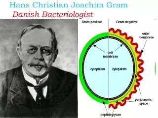
List of the Demonstrated Methods • Haematoxylin & Eosin • v Gieson-elastics • blue trichrom • mucicarmine • PAS • Fe • Kongo red • oil red • impregnation Warthin-Stary ( - Helicobacter) • Gram • Ziehl –Nielsen • polychrom • May-Grünwald-Giemsa