Download
1 / 24
280 likes | 753 Views
GRAM STAINING & ACID FAST STAINING. Dr. R. Haritha Lecturer in Biotechnology Visakha Government Degree College for Women Visakhapatnam. Stains Principle Types Staining methods. Staining. Smear- Distribution of Bacterial cells on a slide

E N D
GRAM STAINING & ACID FAST STAINING Dr. R. Haritha Lecturer in Biotechnology Visakha Government Degree College for Women Visakhapatnam
Stains • Principle • Types • Staining methods Staining
Smear-Distribution of Bacterial cells on a slide Objective-To kill the microorganism & fix bacteria Method- Air Dry, Heat Fixation Smear Preparation
Gram staining is most widely Hans Christian Joachim Gram used differential staining in Microbiology. • It classifies bacteria into two major groups: Gram positive Gram negative Appears violet Appears red after Gram’s stain after Gram’s stain GRAM STAINING
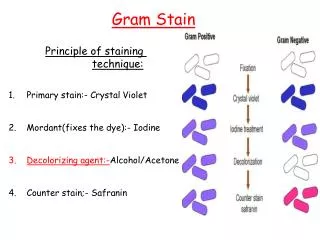
CRYSTAL VIOLET • Primary stain • Violet colored, stains all micro-organism • GRAM IODINE • Mordant • Forms Crystal violet iodine complexes • DECOLORIZER • Acetone + Methanol • Removes Crystal violet iodine complex from thin peptidoglycan layers • GRAM SAFRANINE • Counter stain • Red colored REAGENTS
Step 1:Crystal Violet Step 2:Gram’s Iodine Step 3: Decolorization (Aceton-Alcohol) Step 4:Safranin Red
Gram-positive bacteria • Cell wall has a thick peptidoglycan layer • The Crystal Violet stain gets trapped into this layer and the bacteria turned purple. • Retains the color of the primary stain (crystal violet) after decolorization with alcohol. Gram-negative bacteria • Cell wall has a thin peptidoglycan layer that does not retain crystal violet stain. • Cell wall has a thick lipid layer which dissolves easily upon decoulorization with Aceton-Alcohol. • Therefore, cells will be counterstained with safranin and turned red. PRINCIPLE : Composition of the cell wall
Gram’s +ve Bacteria Gram’s -ve Bacteria
Bacteria that manage to keep the original purple are called Gram positive. • Bacteria that lose the original purple dye and can therefore take up the second red dye are called Gram negative RESULT
1.Rapid preliminary diagnosis of diseases such as Bacterial meningitis. • 2.Selection of antibiotics based on Gram stain finding. • 3.Selection of suitable culture media based on Gram stain finding. • 4.Screening of the quality of the clinical specimens such as sputum that should contain many pus cells & few epithelial cells. • 5.Counting of bacteria. • 6.Appreciation of morphology & types of bacteria in clinical specimens. APPLICATIONS
The Ziehl-Neelsen staining technique. • The acid-fast stain is performed on samples to demonstrate the characteristic of acid fastness in certain bacteria. • Used to identify specialized bacteria that have waxy mycolic acid in their cell wall- MYCOBACTERIA and NOCARDIA ACID FAST STAINING Franz Ziehl Friedrich Neelsen
Due to large quantities of unsaponifiable wax fraction called mycolic acid in their cell wall and also the intactness of the cell wall, these bacteria are highly resistant to staining and treatment. • Heat is used to help drive the primary stain into the waxy cell walls of these difficult-to-stain cells - “Hot Staining” method. • Intense decolonization does not release primary stain from the cell wall of AFB • Color of AFB based on primary stain • Counterstain provides contrasting background PRINCIPLE
Contain large amount of fatty waxes (mycolic acid) within their cell wall • Resist staining by ordinary methods • Require a special stain for diagnostic Acid Fast stain
1. Primary And Mordant Staining with Strong Carbolfuchsin (Red) • 2. Decolourization with Acid Alcohol : The acid alcohol contains 3% HCl and 95% ethanol or 20% H2 SO4. • 3. Counterstain with Methylene Blue. • Acid - Fast Cells Red • Non Acid - Fast Blue REAGENTS
The pink coloured rod shaped structures with curved ends are acid fast bacilli along with the blue coloured pus cells. • The smear is positive for acid fast bacilli. RESULT
A • Teichoic acid C • Peptidoglycan • Peptidoglycan D • Periplasm B • Membrane lipids In Gram Staining Technique, Alcohol acts on.....
Mordant C A Mordant • Fixative • Solubilizer B D Stain In Gram Staining Technique, Iodine is used as.....
A • Crystal Violet C • Saffranin • Saffranin D • Methylene Blue B • CarboIFuchsin The counter stain used in Gram Staining is.....
Mycolic acids C A Mycolic acids • Proteins • Peptidoglycan B D LPS The cell wall of Mycobacterium is made up of.......
C A Acetate • Formaldehyde Alcohol + Acid • Ethyl Alcohol B D Alcohol+Acid The decolourizer used in Acid Fast Staining is...........