Gram Staining Method
Gram Staining Method. Biology 2 Mr. Beyer. Individual Supplies. Microscope Slide Clothes Pin Inoculating Loop or Wood Splint Paper Towels. Shared Supplies. Crystal Violet Stain Safranin Red Stain Gram’s Iodine Ethyl Alcohol (denatured) Water Bottle Rinsing Basin Alcohol Burner

Gram Staining Method
E N D
Presentation Transcript
Gram Staining Method Biology 2 Mr. Beyer
Individual Supplies • Microscope Slide • Clothes Pin • Inoculating Loop or Wood Splint • Paper Towels
Shared Supplies • Crystal Violet Stain • Safranin Red Stain • Gram’s Iodine • Ethyl Alcohol (denatured) • Water Bottle • Rinsing Basin • Alcohol Burner • Wax Pencil • Matches • Microscope
Create a Staining Area On Your Microscope Slide • Obtain a clean microscope slide • Draw a circle on your slide with wax pencil • Warming the slide will make the wax pencil work better when drawing your circle
Create a Suspension of Bacteria • Place 1-2 drops of water inside the circle on your microscope slide • Use the inoculating loop to obtain a tiny sample of bacteria from your bacteria culture • Add the bacteria sample to the water on your microscope slide, mixing to create a suspension of bacteria
Heat Fixing Bacteria to the Microscope Slide • Pass your microscope slide through the flame until all of the water evaporates • This will leave your bacteria stuck or “fixed” to the microscope slide
Primary Staining of Gram Positive Bacteria • Attach the clothes pin to one end of your slide • Place 2-3 drops of Crystal Violet Primary Stain inside your circle (on your bacteria) • Leave the stain on the bacteria sample for one minute • Rinse stain off with water (gently)
Mordant (Sealer) • Add the Gram’s Iodine to your bacteria sample inside the circle • Leave Gram’s Iodine on your sample for one minute • Rinse off with water
De-Colorizing • Add ethyl alcohol to the bacteria • Let the alcohol set, then rinse with water • Continue de-colorizing until the crystal violet stain is no longer released from your sample. • You should just be able to see a hint of purple at this point
Counter-Staining Gram Negative Bacteria • Add 2-3 drops of Safranin Red Stain to your bacteria sample inside the circle on your slide • Leave safranin red on your bacteria sample for one minute • Rinse with water, blot or air dry
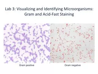
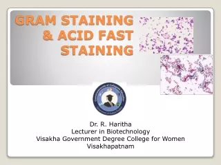
Viewing Bacteria • View under oil immersion lens of microscope (1000x magnification) • Sketch in color, • Label bacteria as gram (+) or gram (-) • Identify shape and arrangement of bacteria