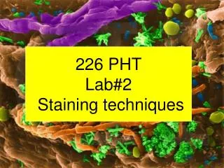
Gram staining Techniques
Gram staining Techniques. Some history. Bacteria are translucent Staining make them visible under microscope 1884 Hans Christian Gram stained cells and found that some lost their color when excess stain was washed off Differential stain distinction between 2 types of bacteria.

Gram staining Techniques
E N D
Presentation Transcript
Some history • Bacteria are translucent • Staining make them visible under microscope • 1884 Hans Christian Gram stained cells and found that some lost their color when excess stain was washed off • Differential stain distinction between 2 types of bacteria http://www2.bvs.org.ve/img/fbpe/rsvm/v23n2/Image140.jpg
Bacteria cell walls • Peptidoglycan: network of sugars cross-linked by short peptides • Forms the rigid part of the cell wall • Protects the bacteria against mechanical damage • Part that picks up the stain in the gram procedure www-micro.msb.le.ac.uk/ video/graphics/wall.gif
Gram - bacteria • Small peptidoglycan • Are stained with crystal violet but decolorized with alcohol after which they pick up the red stain • LPS on outer membrane toxic for host http://www.cat.cc.md.us/courses/bio141/lecguide/unit1/prostruct/toll/u1fig10b.html
Gram + bacteria • Large, highly cross-linked petidoglycan • No outer membrane http://www.cat.cc.md.us/courses/bio141/lecguide/unit1/prostruct/u1fig9b.html
Bacteria shape • Long rod: bacillus • Round shaped: cocci • Spiral: Spirochete
Bacteria shapes http://ibri.org/RRs/RR051/51bacterialshapes-Merc.gif
Some bacteria shapes spirochete bacillus coccobacillus cocci http://www.spcollege.edu/hec/vt/VTDE/ATE2639LGS/images/image11.jpg http://textbookofbacteriology.net/B.anthracis.Gram.CDC.jpeg http://www.furetti.com/images/spirochete%20leptospirosi%201.jpg
Gram stain • Is the most commonly used technique to stain bacteria • Almost always first step into identification • Tell what other tests to perform • Gram – bacteria stain red-pink • Gram + bacteria stain blue-purple
Gram Positive Bacteria (blue/purple) Gram Negative Bacteria (Red/pink)
Gram stain recipe • Smear • Heat fix • Crystal violet (30 sec) wash • Iodine (1 minute) wash • Ethanol (varies ~15 sec) wash • Safranin (30 seconds) wash • Blot dry
How to prepare a smear • Label your slide with a pencil • Use a clean slide: manipulate by the edges • Using a sterilized loop spread of drop of bacterial culture on the slide • If solid culture put a drop of water on the slide add a little bit of bacteria (using loop) • A thin film is better • Dry faster, distribution of dye and decolorizer more even • No coverslip on! let dry (until most water evaporated)
Fixation • Pass the slide through the flame of the bunsen burner • Moving in a circular motion (to avoid localized overheating) • The slide should not be too hot to touch, slightly warm but no more! • If too hot overfixed burn bacteria everything will be black • If underfixed bacteria do not stick to slide and will be washed away during the staining procedure
Why bacteria need to be fixed • Denature bacterial enzyme prevent them from digesting the cell (autolysis) • Make the bacteria stick to the slide so they are not washed away during staining.
1st stain • Crystal violet • Colorize all cells • Flood the slide with crystal violet • ***make sure that the bacteria are on top • 30 seconds • Rinse gently with running tap (or distilled) water • Do not squirt water directly onto the smear • Shake off the excess water
Mordant • Iodine • Make the dye stick to the cell wall • Crystallize the dye in the peptidoglycan • Flood the slide for 1 minute • Wash
Decolorization • Ethanol 15-30 seconds, until dye doesn’t run out anymore • Critical step***** Washing after is very important (stop alcohol action) • Cell that have thin peptidoglycan decolorize • Long story: • Dissolve the lipid layer from the gram negative bacteria • Enhance the leaching from the primary stain • In gram + bacteria: alcohol dehydrate the thicker cell wall • Prevent diffusion of the violet iodine complex
Ethanol Sink
Counter stain • Safranin • color pink • Flood the slide for 30 seconds • Rinse gently with water and shake off the excess water from surface
Blotting • Slides can be air dried or blotted • Blotting put between 2 sheets of absorbent paper (we will use bibulous paper). • DO NOT RUB THE SMEAR (bacteria will come off)…just blot between papers.
Oil immersion • Light is refracted when goes trough slide (change in media from glass to air) • Oil has same refractive index than glass light ray goes with no refraction • Use only one drop of oil • Only use the 40X objective (greatest power objective) with oil immersion. • Clean the objective with paper lens after use
Oil immersion http://www.bmb.psu.edu/courses/micro107/microscopy/oil-lens.jpg
Gram Staining Video • http://www.youtube.com/watch?v=YvZHHNZ8cdo • http://www.bio.upenn.edu/computing/media/Instructional.Stain.Gram.php
Objectives for Tomorrow’s Lab • You have 2 bacteria • S. Urea • P. fluorescens For each bacteria: • Stain each bacteria • Observe and identify the bacteria shape and gram results. • You are doing this lab in team but make sure that each student perform at least 1 gram stain
Resources • www.microvet.arizona.edu/Courses/MIC205/lab/Lab_3_Gram_stain_spring_07.ppt • www.google.com/images/