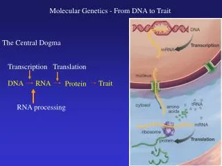
DNA
260 likes | 445 Views
DNA. The Discovery, Replication, DNA vs. RNA. Hershey & Chase. In 1952, American biologists Alfred Hershey and Martha Chase set out to determine what composed the genetic material of a bacteriophage .

DNA
E N D
Presentation Transcript
DNA The Discovery, Replication, DNA vs. RNA
Hershey & Chase • In 1952, American biologists Alfred Hershey and Martha Chase set out to determine what composed the genetic material of a bacteriophage. • They knew that a bacterial virus was an extremely simple organism, composed only of protein and DNA. The protein makes up the exterior of the virus, and the DNA is contained within it.
Hershey & Chase • When a bacterium is infected by a bacteriophage, the bacterium's internal machinery falls under the control of the virus, which uses the bacterium to produce more viruses. • What Hershey and Chase wanted to know was: Which substance directed this takeover - DNA or protein?
Hershey & Chase • The Experiment: • Protein contains sulfur. DNA contains phosphorus. • They added bacteriophage (a bacterial virus) to cultures containing either radioactive sulfur (S35) or radioactive phosphorus (P32). • Hershey and Chase now had two types of bacteriophages: one with a radioactive external protein coat, the other with radioactive DNA.
Hershey & Chase • The bacteriophages were allowed to infect the bacteria. The phage injects its DNA into the bacterial cell, while the protein coat remains outside. • Using methods to separate the liquid and used phage protein coats from the bacterial cells… • In the cultures infected by bacteriophages with radioactive sulfur, the radioactivity was in the liquid with the phages.
Hershey & Chase • In the cultures infected by bacteriophages with radioactive phosphorus, the radioactivity was in the pellet of infected bacteria. • Thus, Hershey and Chase discovered that the radioactive protein hadn't entered the bacterial cells, but the DNA had. • Lets take a look: http://highered.mcgraw-hill.com/olcweb/cgi/pluginpop.cgi?it=swf::535::535::/sites/dl/free/0072437316/120076/bio21.swf::Hershey%20and%20Chase%20Experiment
Wilkins &Franklin • Maurice Wilkins shared the 1962 Nobel Prize for Physiology and Medicine with Watson and Crick for the discovery of the structure of DNA. • Why was Rosalind Franklin not honored for her truly essential contribution to this discovery, and could not be recognized by the Nobel Committee in 1962?
Wilkins &Franklin • In 1952 Maurice Wilkins and Rosalind Franklin were given the task of determining the molecular structure of DNA. • They investigated DNA through the scientific technique of X-ray crystallography, in which Rosalind was very skilled. • She took the pictures and made the calculations that led to the discovery of the shape of DNA.
Wilkins &Franklin • Rosalind Franklin’s X-ray diffraction Photo 51 (B) which clarified the double helix shape of DNA.
Watson & Crick • In 1953, Francis Crick and James Watson described the structure of DNA. • Their model of DNA was based on their own DNA research AND the research of several other scientists including: • Linus Pauling, 1948, who discovered that many proteins take the shape of an alpha helix, spiraled like a spring coil.
Watson & Crick • Biochemist Erwin Chargaff, 1950, found that the arrangement of nitrogen bases in DNA varied widely, but the amount of certain bases always occurred in a one-to-one ratio. • Maurice Wilkins and Rosalind Franklin’s __________________photos of DNA double helix, 1952.
DNA Structure • DNA stands for deoxyribonucleic acid. • Nucleotides are the units that make-up DNA. • Nucleotides have three parts: • - 5 Carbon sugar – Deoxyribose • - a phosphate group • - a nitrogen base (one of four) • * arranged along the backbone in 5’ • (phosphate) to 3’ (OH) direction.
DNA Structure • Helical shape, Double-strand pairing, Antiparallel (one strand 5’ to 3’, the other 3’ to 5’) • Four different nitrogen bases: • Adenine, Thymine, Cytosine, Guanine • Complementary base-matching: A-T, C-G • Base-matching achieved by Hydrogen bonding and geometry • A, G are long, double ring purines • T, C are short, single ring pyrimidines
Nucleotide Structure 5’ 5’ 3’ 3’
DNA Replication • DNA replication is semi-conservative. Meaning one strand from each of the initial two parent strands ends up in a new daughter strand. • Each strand serves as a template for a new strand. • New strand is formed by complementary base-pairing of the correct nucleotides.
DNA Replication • DNA replication begins when helicase unwinds a segment of the DNA and breaks the hydrogen bonds between the two complementary strands. • DNA polymerase can only add new nucleotides to a free 3’ end of a growing chain. Synthesis of one strand of the DNA, called the leading strand, proceeds continuously in the 5’ to 3’ direction. • Synthesis of the complementary strand, called the lagging strand, is more complex.
DNA Replication • DNA polymerase can add new nucleotides only to a free 3’ OH end. • To provide a free 3’ OH starting point, RNA primase attaches to the DNA and synthesizes a short RNAprimer. DNA polymerase III then adds new nucleotides to the 3’ end of the RNA primer. (Adds nucleotides between primers.) • DNA polymerase I replaces DNA polymerase III, removes the RNA primer and replaces it with DNA nucleotides.
DNA Replication • DNA ligase forms a phosphodiester bond between the 3’ OH of the growing strand and the 5’ phosphate in front of it. • During DNA replication, the leading strand is synthesized continuously, while the lagging strand is synthesized discontinuously. • Lets take a look: http://highered.mcgraw-hill.com/olcweb/cgi/pluginpop.cgi?it=swf::535::535::/sites/dl/free/0072437316/120076/micro04.swf::DNA%20Replication%20Fork
DNA Replication • The pieces of new DNA strand between the RNA primers are called Okazaki fragments.
DNA vs. RNA • RNA • DNA Make yourself a chart!
DNA vs. RNA • RNA • Ribose sugar • DNA • Deoxyribose sugar
DNA vs. RNA • RNA • Ribose sugar • Single Strand • DNA • Deoxyribose sugar • Double Strand
DNA vs. RNA • RNA • Ribose sugar • Single Strand • Bases: A, C, G, U • DNA • Deoxyribose sugar • Double Strand • Bases: A, C, G, T
DNA vs. RNA RNA Ribose sugar Single Strand Bases: A, C, G, U A “Messenger” of information • DNA • Deoxyribose sugar • Double Strand • Bases: A, C, G, T • For the “Storage” of information