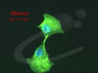
Mitosis
180 likes | 365 Views
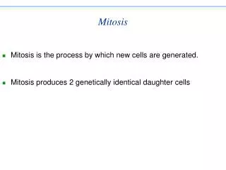
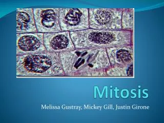
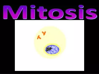
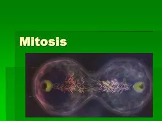
Mitosis. Mitosis in animal cell. Prophase. The two round objects above the nucleus are the centrosomes . The chromatin is condensing into chromosomes. Prometaphase.

Mitosis
E N D
Presentation Transcript
Mitosis in animal cell Prophase The two round objects above the nucleus are the centrosomes. The chromatin is condensing into chromosomes. Prometaphase The nuclear membrane disintegrates, and microtubules have invaded the nuclear space. These microtubules can attach to kinetochores or they can interact with opposing microtubules. Metaphase The chromosomes align at the metaphase plate. The chromosomes split and the kinetochore microtubules shorten. Anaphase Telophase The decondensing chromosomes are surrounded by nuclear membranes. Cytokinesis has already begun; the pinched area is known as the cleavage furrow (cell plate).
Germanbiologist and a founder of cytogenetic Drawing of mitosis by Walther Flemming.
Meiosis • The form of cell divisionby which gametes, with half the number of chromosomes,are produced. • Diploid (2n) haploid (n) • Meiosis is sexual reproduction. • Two divisions (meiosis I and meiosis II).
Meiosis • Sex cells divide to produce gametes(sperm or egg). • Gametes have half the # of chromosomes. • Occurs only in gonads (testes or ovaries). Male: spermatogenesis Female: oogenesis • Meiosis is similar to mitosis with some chromosomal differences.
Meiosis – key differences from mitosis • Meiosis reduces the number of chromosomes by half. • Daughter cellsdiffer from parent, and each other. • Meiosis involves two divisions, Mitosis only one. • Meiosis I involves: • Synapsis – homologous chromosomes pair up. Chiasmata form (crossing over of non-sister chromatids). • In Metaphase I, homologous pairs line up at metaphase plate. • In Anaphase I, sister chromatids do NOT separate. • Overall, separation of homologous pairs of chromosomes, rather than sister chromatids of individual chromosome.
Interphase I • Similar to mitosis interphase. • Chromosomes replicate (S phase). • Each duplicated chromosome consist of two identical sister chromatids attached at their centromeres. • Centriole pairs also replicate.
Meiosis I (four phases) • Cell division that reduces the chromosome number by one-half. • four phases: a. prophase I b. metaphase I c. anaphase I d. telophase I
Leptotene Zygotene Pachytene Diplotene Diakinese Anaphase -II Anaphase -I Telophase-I Metaphase -II Metaphase -I Telophase-II
Leptotene(Gr. leptos = thin; taenia = band / stripe) Leptoteneis the first stage of meiosis. This is also the first step in the condensation of DNA, a phenomenon that proceeds through the entire prophase I. Despite the thread-like aspect of th chromosomes each consist in fact of twee chromatides, because the DNA has been already replicated during the premeiotic S-fase. Also small regions with thickenings (so-called chromomeres) arise in the chromatine on each chromosome, which make them look like a pearl nacklace. The homolog chromosomes are still unpaired. Zygotene(Gr. zygon = touching another) Zygotene (Gr. zygon = touching another), the stage after leptotene, the pairing of the homolog chromosomes (synapsis) begins. The homolog chromosomes are hold together by proteins (synaptomal complex). Next, crossing-over can happen between the DNA-double helices molecules of homolog chromosomes.
Pachytene(Gr. pachus = dik) Pachytene, a stage of prophase I, are the complete pairing of the chromosomes and the lining-up of the chromomeres. Synapsis, that commonly shifts like a zip from the telomeres to the centromeres, is now at a climax. The nucleoli are often still visible at pachytene. Diplotene (Gr. diplous = in twofold) During diplotene, it becomes clearly visible that each replicated chromosome consists of two sister-chromatides. Each bivalent consist of a bundle of four homolog chromatides. Homologs exhibit a weaker binding and slightly diverge. The crossings (= chiasmata; singular chiasma, named after the cross-shaped lgreek letter chi) between non-sister-chromatides are visible. Each bivalent shows in general one or more chiasmata, where crossing-overs have occured.
Diakinesis(Gr. dia = apart; Gr. kinein = to move) At diakinesis, the chromatides that we in a crossing-over are entangling. Since the chromatidesdeiverge the chiasmata become clearly apparent. Nuclear membrane and nucleoli completely disappear. The spindle arises: from each centrosomes (in animal cells) at the poles microtubules 'grow'. Some microtubules anchor to the kinetochores of the chromosomes. The shortening and thickening of the chromatides proceeds. Metaphase I (Gr.meta=in the midth) At metaphase I (Gr.meta=in the midth) the nuclear envelop and nucleoli have now disappeared and each pair of homolog chromosomes moves to the equatorial plane, with the centromeres protruding at each side of this plane. The two centromeres of any homolog pair of chromosomes are attached each to spindle microtubules reaching opposite poles Equatorial plane Polar view.
Anaphase I The chromosomes move apart toward the opposite poles. At this stage of meiosis I the centromeres do not separate: chromatides within one chromosome stay together. This is a very important DIFFERENCE with anaphase in mitosis TelophaseI and the consecutive interphase (interkinesis) do not generally occuring in samples of meiottic divisions. In many organismd these phase are even skipped; this implies that no new nuclear membrane is built around the two nuclei between after anaphase I and that the cell directly proceeds to meiosis II. In other organisms telophase I and interkinesis last very shortly; the chromosome temporarily despiralize and are less visible for a period, while a nuclear membrane is formed around each new nucleus. What ever the scenario is prefered in this intermediate phase there occurs NEVER doubling of the DNA, not does genetic reshuffling take place, thus also no crossing-over among chomosomes Telophase I (Gr. telos = end)
Prophase II This stage at the beginning of meiosis II is characterized by the presence of a haploid number of chromosomes that condense again. Metaphase II The chromosomes move again to the equatorial plane between the poles. However, this plane is perpendicular to the equatorial plan of Metaphase I.
Anaphase I The centromeres separate and the chromatides are pulled by the spindle microtubules to opposite poles (perpendicular to the axis of anaphase I) Telophase II By the end of meiosis II a new nuclear envelop is formed around each of the four new nuclei, while the chromosomes despiralize. Tetrad Tetrad (Gr tetra = four) is the name given to the period within post-meiostic differentiation during which the four microspores (sometimes also the megaspores) are still together. They are only separated by a temporary cell wall.