Download
1 / 47
480 likes | 647 Views
Warm Up. I’m passing back your quizzes, please review your grade carefully (there were many mistakes on Pd 6’s papers) Also be sure you were not marked wrong for questions 5&6 MC and 4 T/F Raise your hand if there is a grading issue and I will fix it Test corrections are due on Monday.

E N D
Warm Up • I’m passing back your quizzes, please review your grade carefully • (there were many mistakes on Pd 6’s papers) • Also be sure you were not marked wrong for questions 5&6 MC and 4 T/F • Raise your hand if there is a grading issue and I will fix it • Test corrections are due on Monday
Video: Tree Man FINALLY!
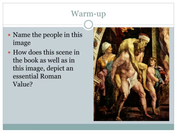
Warm Up On a lined sheet of paper: explain tree man’s disease. What is it caused by? How can it be treated?
Intro to Bones Do we know any bones in the body? Video
Notes: Skeletal System! • Skeletal System Overview: • Axial skeleton • Appendicular skeleton • Joints • Human body is made up of 206 bones!
Function of Bone • Support • provides internal framework • Protection • surround vital organs • Movement • attached to tendons attached to muscles • Storage • contains minerals (calcium) and stores fat in bone marrow • RBC formation • in bone marrow
Classification of Bones • Types of Bone: • Compact—dense, smooth, homogenous • Spongy—porous • Classification of Bones: • Long bones—long shaft with heads at both ends that is mostly compact bone • EX: appendages (except kneecap, wrist, and ankle) • Short bone—cube-shaped that is mostly spongy bone • EX: kneecap (patella), wrist, ankle) • Flat bones—thin, flat, curved that is two thin layers of compact bone sandwiching one layer of spongy bone • EX: skull, ribs, sternum • Irregular bones—all others • EX: vertebrae, hip bones
Anatomy of Long Bones • Haversian canals • run lengthwise carrying blood vessels and nerves • Lacunae • cavities of bones cells (osteocytes) • Lamellae • concentric circles of lacunae around one Haversian canal • Osteon • one unit consisting of one Haversian canal and accompanying lacunae/lamallae • Canaliculi • run widthwise in each osteon carrying nutrients • Volkmann’s canals • run widthwise between osteons carrying blood vessels and nerves
Group Work: Anatomy of Long Bone Read the front of the handout Then, color the back of the handout with colored pencils
Warm Up Name the two types of bone based on their makeup. Also, take out your coloring homework for Ms. McGowan to check
Gross Anatomy of Long Bones • Diaphysis—shaft of long bone • Periosteum—fibrous connective tissue membrane that covers diaphysis • Perforating fibers—aka Sharpey’s fibers, secure periosteum to diaphysis • Epiphyses—heads/ends of long bone, thin layer of compact bone enclosing spongy bone • Articular cartilage—glassy hyaline cartilage membrane that covers epiphyses • Ephiphyseal plate—causes lengthwise growth until inhibited by hormones at end of puberty • Ephiphyseal line—bone that replaces epiphyseal plates post puberty • Yellow marrow—cavity of shaft filled with adipose tissue (in adults) • Red marrow—cavity of shaft that forms RBCs (in children) • RBCs formation in adults only in flat bones and epiphyses
Label: Left Coronal suture Parietal bone Temporal bone Lambdoid suture Squamous suture Occipital bone Mandibularramus Right Frontal bone Nasal bone Zygomatic bone Maxilla Alveolar margins Mandible
Label: Left Foramen magnum Right Cribriform plate
Label: Left Hard palate, etc. Vomer Right Foramen magnum
Label: Left Coronal suture Parietal bone Nasal bone Zygomatic bone Maxilla Mandible Right Frontal bone Temporal bone Vomer Alveolar margins
Label: Hyoid bone
Warm Up Sketch the skull below Label the 4 major bones of the skull Turn in test corrections!
Group Work: Labeling Skull With a partner, label your skull model in pen or marker with the terms you just learned
Notes: Vertebral Column and Thoracic Cage Vertebral Column • Aka spine • Connected to skull and pelvis, forming a curved S shape • Protects spinal cord that runs within
Made of 26 irregular bones (from top to bottom): • Cervical spine (C1-C7) • Think breakfast at 7am! • Smallest and lightest, characteristic foramen in transverse processes • Thoracic spine (T1-T12) • Think lunch at 12pm! • Larger than C spine, only vertebrae to connect to ribs • Lumbar spine (L1-L5) • Think dinner at 5pm! • Massive bodies to support majority of weight • Sacrum • 5 fused vertebrae • Forms posterior wall of pelvis • Coccyx • 4 fused vertebrae • Aka tailbone
Warm Up What are the 5 portions of the spine? How will you remember how many vertebrae are in the main three portions?
Vertebrae connected by flexible intevertebral discs that cushion and absorb shock • General vertebrae characteristics: • Body or centrum—disc like, weight-bearing part of the vertebra facing anteriorly in the vertebral column • Vertebral arch—joins laminae and pedicles • Vertebral foramen—canal through which spinal cord passes • Transverse processes—two lateral projections • Spinous process—single medial, posterior projection • Superior and inferior articular processes—paired projections lateral to the vertebral foramen, allows formation of joints between vertebrae
Thoracic Cage • Protects heart and lungs • Thoracic spine (see above) • Sternum • Aka breastbone • Fusion of 3 bones (from top to bottom): • Manubrium • Body • Xiphoid process • Attached to first 7 pairs of ribs • 3 landmarks • Jugular notch—concave upper border of manubrium at level of T3 • Sternal angle—where manubrium and body meet at angle, forming ridge at level of 2nd ribs • Xiphisternal joint—where body and xiphoid meet at level of T9
Ribs • 12 pairs that all connect posteriorly with vertebrae • True ribs—first 7 pairs, attach directly to sternum by cartilage • False ribs—next 5 pairs, attach indirectly to sternum through much cartilage • Floating ribs—last 2 pairs, not connected to cartilage or sternum • Intercostal spaces between ribs filled with intercostal muscles that aid in breathing
Group Work: Review Work together, to complete your review sheet
Warm Up While at a lacrosse game, your friend was checked very hard in the ribs. When you ask him if he is ok, he responds, “Yea, bud he only hit me in my fake ribs—you know, the ones we don’t need?” How would you respond to your friend?
Closing HAPPY HOMECOMING!
Warm Up Any last questions?
Axial Skeleton Quiz! Clear your desk except for a writing utensil If you have a question, raise your hand When you finish, hold on to your quiz and put your head down—I will collect all quizzes at the end of class Good luck!
Closing How was it?
Warm Up How was the quiz?
Microscopic Anatomy of Long Bone Checkpoint Observe a section of long bone under the microscope Label the microscopic anatomy of long bone using learned terminology
Guided Notes: Appendicular Skeleton The three parts of the appendicular skeleton are the: • Limbs • Pectoral girdle • Pelvic girdle
Guided Notes: Appendicular Skeleton • Pectoral (Shoulder) Girdle • Light and free to movement because only attached to axial skeleton at sternoclavicular joint • But very easily dislocated! • Two parts: • Clavicle—collarbone • Attached to manubrium and scapulae • Holds arm away from top of thorax and prevents shoulder dislocation • Scapulae—shoulder blade • Not directly attached to axial skeleton • Attached to clavicle and trunk muscles • Anchors arm muscles
Warm Up What three parts make up the appendicular skeleton?
Guided Notes: Appendicular Skeleton • Upper Limbs • Arm--humerus • Rounded head connects to scapula • Non-rounded head connects to radius • Surgical neck—common site of fracture • Forearm • Two parts: • Radius • On thumb side • Ulna • On pinky side • Connected at radioulnar joints • Also connected along entire length by flexible interosseous membrane
Guided Notes: Appendicular Skeleton • Hand • Three parts: • Carpals--wrist • 8 bones; 2 irregular rows of 4 bones each • Bound together by ligaments that restrict movement between them • Metacarpals--palm • Numbered 1-5 from the thumb side • When you clench your fist, the knuckles you observe are the heads of the metacarpals • Phalanges--fingers • 14 bones; 3 bones for each finger (labeled proximal, middle, distal) except 2 bones for the thumb
Group Work: Pelvis In your groups, analyze the two pelvis photos given Then, write down as many differences between the two as you can
Guided Notes: Appendicular Skeleton • Pelvic Girdle • Two parts: • Two coxal (hip) bones • Sacrum • (Not to be confused with pelvis which is made up of pelvic girdle plus coccyx) • Attached to axial skeleton via sacrum • Large and heavy bones • Bear all upper body weight • Also protect reproductive organs, bladder, and part of colon
Guided Notes: Appendicular Skeleton • Hip bone • Three parts: • Ilium • Makes up majority of hip, most superior portion • When rest hands on hip, resting on ilium • Connected to sacrum • Iliac crest—top of ilium, landmark for intermuscular injections • Pubis • Inferior to ilium, anterior to ischium • Combines with ischium to make obturator foramen • Ischium • Posterior to pubis • Ischialtuberosity--inferior portion that receives body weight while sitting • Ischial spine--superior to ischialtuberosity that narrows canal through which baby passes during birth • Greater sciatic notch--notch through which sciatic nerve passes from spine to thigh
Guided Notes: Appendicular Skeleton • General • Pubic symphysis—area where both coxal bones fuse inferiorly and medially • Pubic arch—shape formed directly inferior to pubic symphysis • Acetabulum—area where ilium, pubis, and ischium fuse • also site where head of femur connects to hip • False pelvis—superior, between flaring portions of ilia • True pelvis—inferior, below flaring portions of ilia • Size of true pelvis needs to be large enough for childbirth
Guided Notes: Appendicular Skeleton • Men vs. Women • Female true pelvis larger and more circular • Female pelvis shallower and the bones are lighter and thinner • Female ilia flare more laterally • Female sacrum shorter and less curved • Female ischial spines shorter and farther apart; thus false pelvis is larger • Female pubic arch more rounded because the angle of the pubic arch is greater