Download
1 / 73
730 likes | 750 Views
Learn about the different senses and their receptors, explore the two-point threshold, and understand the anatomy of the eye.

E N D
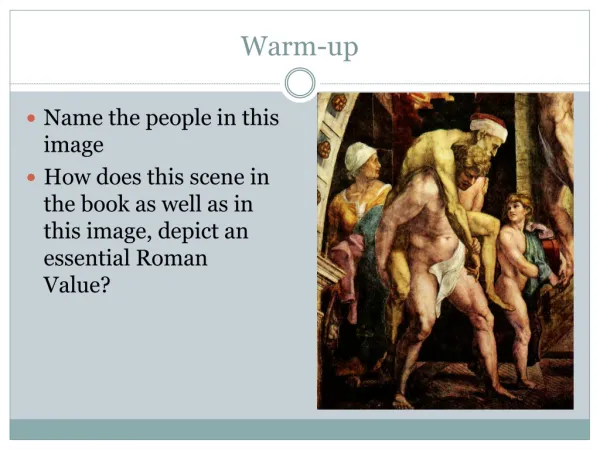
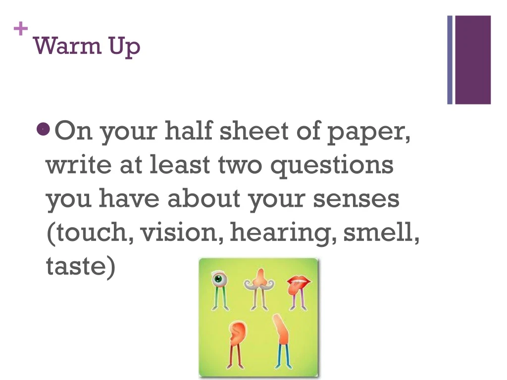
Warm Up • On your half sheet of paper, write at least two questions you have about your senses (touch, vision, hearing, smell, taste)
Objective: • SWBAT • Explain why certain areas of the body are variously sensitive and therefore have different sized receptive fields • Agenda: • Notes: Intro to Senses and Touch • Do you get the point? • Closing • Homework: • Finish Lab
Guided Notes: Intro to Senses • Sensation—the conscious or subconscious awareness of the internal and external conditions of the body • Detected by receptors • Type of receptor varies depending on the sense • Mechanoreceptors—responds to mechanical stimuli for touch, stretch, pressure, position, and hearing • Meissner’s corpuscles and Merkel disks—touch • Pacinian corpuscles--pressure • Proprioceptors—position • Thermoreceptors--temperature • Nocioceptors—pain • Photoreceptors—responds to light stimuli for vision • Chemoreceptors—responds to chemical stimuli for smell and taste
Guided Notes: Intro to Senses • 4 steps of each receptor • Reception—detection of stimuli by sensory receptors • Transduction—conversion of stimuli into change in membrane potential of sensory receptor • Transmission—if threshold is reached, action potential propagates to the brain • Integration—brain’s construction of stimuli • Amplification—strengthening of stimulus by exciting more neurons • Adaptation—weakening of stimulus due to continued stimulation • To prevent cell death • Fast adapters—thermoreceptors, pressure receptors, and touch receptors • Slow adapters—nocioceptors and proprioceptors
Guided Notes: Touch • Receptive field—skin area served by a single sensory neuron • Inversely proportional to the density of sensory receptors (sensitivity) • Two point touch threshold—the minimum distance at which the touch of two points can be distinguished • Proportional to the size of the receptive field • Inversely proportional to the density of sensory receptors (sensitivity) • Referred pain—presence of pain felt in part of body different from part of body that is actually generating pain • Due to convergence of sensory receptors • EX: heart and left upper limb enter spinal cord at same level • EX: brain freeze • EX: Phantom Limb—perception of stimuli at location of amputated limb
Do You Get the Point? • Working with a partner, explore the two point threshold of your forehead, cheek, back of forearm, palm of hand, tip of thumb, tip of index finger, and back of lower leg using two paperclips
Warm Up • Why would a neurologist administer a two point discrimination test on a patient?
Objective: • SWBAT • Explain why certain areas of the body are variously sensitive and therefore have different sized receptive fields • Agenda: • Finish Do you get the point? • Go over Do you get the point? • Closing • Homework: • None!
Do You Get the Point? • Working with a partner, explore the two point threshold of your forehead, cheek, back of forearm, palm of hand, tip of thumb, tip of index finger, and back of lower leg using two paperclips
Warm Up • What part of your body had the smallest two point threshold? • Why do you think this is? • What does this mean in terms of size of receptive field, density of neurons, and sensitivity? • What part of your body had the largest two point threshold? • Why do you think this is? • What does this mean in terms of size of receptive field, density of neurons, and sensitivity?
Objective: • SWBAT • Identify at least ten major structures of the eyeball • Explain why we become congested after crying • Agenda: • Last call for “Do You Get the Point?” Lab • Guided Notes: Anatomy of the Eye • Coloring and Labeling • Application Questions • Homework: • Finish Application Questions
Guided Notes: Anatomy of the Eye • Internal Structures • Sclera—outermost connective tissue layer that surrounds eye everywhere except cornea • Reason we see whites of eyes • Cornea—clear covering over visible portion of eye • where light enters eye • Contains nocioceptors because cornea very vulnerable to damage • Easily repairs self • No blood vessels so easily transplanted with no fear of rejection • Choroid—vascular layer under sclera • Ciliary muscle and fibers—smooth muscle and fibers at anterior of eye attached to the lens and the iris • Iris—pigmented smooth muscle that regulates amount of light entering eye • Gives eye color • Constrict for bright light or close vision • Dilate for dim light or distant vision • Pupil—round opening in iris through which light passes
Retina—innermost sensory layer under choroid that only extends posteriorly from ciliary body and lens • Photoreceptors • Rods—responsible for gray tones in dim light, concentrated peripherally • Cones—responsible for color, concentrated centrally, red, green, and blue cones—if all stimulated, we see white • Signal moves from photoreceptors to bipolar cells to ganglion cells to optic nerve • Blind spot--site where optic nerve leaves eyeball • Fovea—site next to blind spot that only contains cones, area of sharpest vision • Lens—flexible biconvex structure that focuses light on retina • Aqueous humor—clear watery fluid anterior to lens • Provides nutrients for cornea and lens • Maintains intraocular pressure • Vitreous humor—gel-like fluid posterior to lens • Prevents eye from collapsing in on self • Maintains intraocular pressure
Accessory Structures • Eyelids with eyelashes • Conjunctiva—the outermost membrane that secretes mucus to lubricate the eyeball • Lacrimal gland—structure superior and lateral to eyeball that produces salty tears • Tears contain antibodies and lysozymes • Tears move across eyeball medially and drain into nasal cavity • 6 eye muscles
Coloring, Labeling, and Application Questions • Label the eyeball diagram on the back of your notes sheet • Then, color each structure a different color • When you finish coloring, answer you application questions
Warm Up • Why do we become congested after crying? • Pass your Application Questions to the aisle for collection
Objective: • SWBAT • Explain the cause of at least 5 vision problems • Agenda: • Last call for “Do You Get the Point?” Lab • Guided Notes: Vision and Vision Problems • Application Questions • Homework: • Close Reading: Vision
Guided Notes: Vision • Lens Refraction • Refraction—bending of light rays so that they focus at any given point on the retina • Lens is “set” for distance vision • Ciliary muscle is relaxed, so fibers holding lens are taut, lens is flat • Accomodation—ability for lens to change shape for close vision • Ciliary muscle contracts, so fibers holding lens slack, lens rounds
Guided Notes: Vision • Visual Fields and Visual Pathways to the Brain • Optic chiasma—fibers from the medial side of each eye cross over to the opposite side of the brain • Optic tracts—contains fibers from the lateral side of the eye on the same side and the medial side of the opposite eye • Two similar but different images from both eyes • Results in depth perception and 3D images • Integrated in thalamus and occipital lobe
Guided Notes: Vision • Development • Doctor regularly administers fundoscopic exam • Uses ophthalmoscope to visualize fundus—posterior of eye • Will observe retina, blood vessels, and internal disc to determine if DM, arteriosclerosis, or degeneration of optic nerve or retina
Guided Notes: Vision Problems • Eye Issues • Night blindness—prolonged vitamin A deficiency causes deterioration of rods • Color blindness—lack of cones • Most common: sex-linked red-green color blindness in men • Use differences in intensities to determine red from green • Cataracts—hardening of lens resulting in hazy vision and eventually blindness • Risk factors: DM, too much sunlight, smoking • Treatment: replace lens • Glaucoma—blockage of aqueous humor drainage resulting in increase in intraocular pressure and therefore damage to retina and optic nerve • Common cause of blindness in elderly because progresses slowly with no symptoms • To prevent, doctors check intraocular pressure yearly in patients over 40 with tonometer • To treat, eyedrops that increase drainage or laser or surgical enlargement of drain
Guided Notes: Vision Problems • Myopia—“nearsightedness,” can see close objects, distant objects blurry because image focused in front of retina, need concave lenses • Hyperopia—“farsightedness,” can see far objects, close objects blurry because image focused behind retina, need convex lenses • Astigmatism—cornea or lens is more oval shaped resulting in blurred vision • Nystagmus—rapid involuntary eye movement • Strabismus—“cross-eyed” or misalignment of eyes due to cranial nerve damage • Can lead to amblyopia—“lazy eye” because child’s brain will ignore one image over the other • Hemianopia—CVA damages one optic tract, resulting in loss of vision on one side of visual field (left or right depending on site of CVA)
Application Questions • Work together to answer your application questions • When you finish, complete your reading
Warm Up • Why do long periods of reading or computer work or video games result in eyestrain? • Why does periodically staring into the distance prevent and relieve eyestrain? • Pass your application questions and close reading to the aisle for collection
Objective: • SWBAT • Discuss the efficiency of the multitude of tests available to test vision issues such as those associated with acuity, astigmatism, and colorblindness • Agenda: • Collect Application Questions and Close Reading • Vision Lab • Closing • Homework: • Finish Vision Lab Analysis Questions • Wear appropriate clothing for cow eye dissection tomorrow!
Vision Lab • Move around to each station, to complete your lab sheet
Closing • How do did you test for visual acuity during your lab? • Is this the best way to test visual acuity?
Warm Up • How d • Pass your vision lab to the aisle for collection
Objective: • SWBAT • Identify and explain the importance of the major structures of the sheep eye, including the sclera, cornea, choroid, ciliary muscles, ciliary fibers, iris, pupil, retina, blind spot, lens, aqueous humor, and vitreous humor. • Agenda: • Collect Vision Lab • Eye Dissection • Closing • Homework: • None!
Eye Dissection • Working with your lab table, follow the directions on your lab worksheet to dissect the sheep eye
Closing • What surprised you most about this dissection?
Warm Up • Why do animals eyes “glow” in the dark when light is shined on them at night? Consider what this structure is called and what purpose it holds in the animal eye. • Pass your Eye Dissection Lab and Close Reading: Why do limbs fall asleep? to the aisle for collection.
Objective: • SWBAT • Self assess their knowledge of touch and vision informally through review prior to tomorrow’s touch and vision quiz. • Agenda: • Touch and Vision Quiz Review • Brain Games: In Living Color • Closing • Homework: • Touch/Vision Quiz Tomorrow
Touch and Vision Quiz Review • Take out your touch and vision quiz review • Follow along as we go over the answers
Brain Games: In Living Color • As you watch, consider the question, why do we see after images?
Warm Up • Why do we see after images?
Objective: • SWBAT • Assess their knowledge of touch and vision formally through their touch and vision quiz. • Agenda: • Touch and Vision Quiz • Closing • Homework: • None!
Touch and Vision Quiz • Put everything away except for a writing utensil • If you have a question during your quiz, raise your hand and I will come answer it • Hold on to your quiz when you finish; I will collect them all at the same time • Good luck!
Closing • How was your quiz? • Why do you think you performed in this way? • Consider how you participated in class, how you studied, and how Ms. McGowan presented the information
Warm Up • What memory comes to mind when you think of the smell of chocolate chip cookies?
Objective: • SWBAT • Explain why certain people like certain smells while others dislike them • Explain why an individual’s smell preference changes over time • Agenda: • Guided Notes: Smell • Smell Lab • Closing • Homework: • None!
Guided Notes: Smell • Anatomy of Olfactory Receptors • Pathway: • Olfactory hairs (cilia) coated in mucus make up a postage stamp-size area on roof of nasal cavity • Chemicals dissolve into the mucus • Olfactory hairs respond to chemicals by sending action potentials down the olfactory nerve • Olfactory nerves bundle and transmit signals to the olfactory cortex of the brain for integration and interpretation
Guided Notes: Smell • The olfactory pathway is closely tied to the limbic system (emotions and memory) in brain • This is why we like certain smells and dislike certain smells! • This is why our smell preference can change over time! • Chemoreceptors are fast-adapters
Guided Notes: Smell • Development • Highest functioning at birth • During mid 40s, chemoreceptors decrease in number, resulting in smell deficits • Half of those over 80 can’t smell at all • Issues • Anosmia—temporary or permanent loss of smell due to nasal cavity inflammation, head injury, or aging • Epileptics often experience olfactory auras (or hallucinations) preceding seizures
Smell Lab! • Working individually, move around the room to each lab station. • At each lab station, “puff” the fragrance near your nose. • Then, determine your smell preference (whether you like, dislike, or don’t have a preference for the smell). • Next, describe the memory that comes to mind when you smell this fragrance. • Finally, attempt to identify the smell from the 7 options given. • When you are finished, return to your seat and quietly answer your analysis questions. • We will have a class discussion when everyone is done collecting data.
Closing • Why do some people like certain smells while others dislike them? • Why does an individual’s smell preference change over time?
Warm Up • Are senses “connected”? If so, which ones?