Download
1 / 44
450 likes | 618 Views
Chapter 1. Histology: The Study of Tissues. Tissue Level of Organization. The classification of tissue types is based on the structure of cells; the composition of noncellular substance surrounding cells (extracellular matrix) and the functions of the cells. Tissues and Histology.

E N D
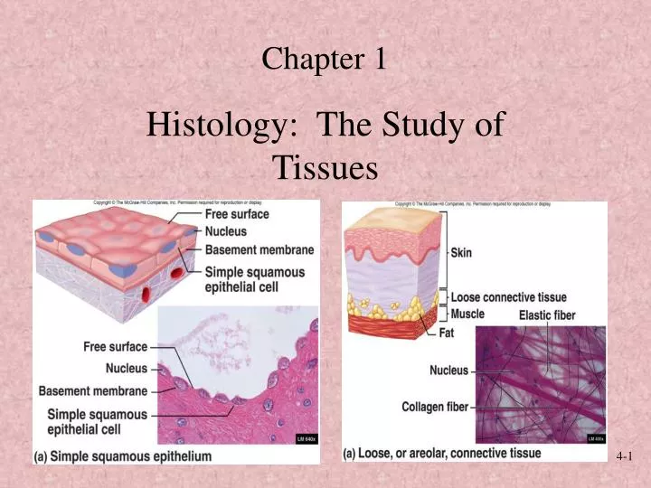
Chapter 1 Histology: The Study of Tissues
Tissue Level of Organization • The classification of tissue types is based on the structure of cells; the composition of noncellular substance surrounding cells (extracellular matrix) and the functions of the cells.
Tissues and Histology • Tissue Level of Organization • Epithelial • Connective • Muscle • Nervous • Histology: Microscopic Study of Tissues
Overview of Tissue Science • An Orientation to the Tissues of the Body Figure 4-1
Epithelium Characteristics • Consists almost entirely of cells • Covers body surfaces and forms glands` • Has free and basal surface • Avascular • No spaces between cells
Functions of Epithelia • Protecting underlying structures • Acting as barriers • Sensation • Secreting substances • Absorbing substances • Excrethion • Diffusion, Permitting the passage of substances • Cleaning • Reduce friction • contraction
Classification Accordingto the: • Shape of cells • No. of layers
Types of Epithelium • Types of epithelium is based on the shape of the epithelial cells: • Squamous: cells are flat • Cuboidal: cells are cube-shaped • Columnar: cells are taller tan they are wide.
Classification of Epithelium • Simple • Squamous, cuboidal, columnar • Consists of a single layer of cells with each extending from the basement membrane to the free surface • Stratified • Squamous, cuboidal, columnar • Consists of more than one layer of cells, only one of which is attached to the basement membrane.
Classification of Epithelium • Pseudostratified • Columnar • Special type of simple epithelium • It appears to be stratified but it is not (false – psuedo) • Consists of one layer of cells, with all the cells attached to the basement membrane.
Classification of Epithelium • Transitional • Cuboidal to columnar when not stretched and squamouslike when stretched
Simple Squamous Epithelium • Consists of a single layer of cells, with each cell extending from the basement membrane to the free surface.
Simple Squamous Epithelium • Function: diffusion, filtration, some protection against friction, secretion and absorption • Location:lining of blood and lymphatic vessels (endothelium) and small ducts, alveoli of the lungs, loop of Henle in kidney tubules,line the heart.
Simple Cuboidal Epithelium • Cuboidal: cells are cube-shape; about as wide as they are tall. • Single layer of cube-shaped cells, some cells have microvilli or cilia.
Simple Cuboidal Epithelium Figure 4-4(b)
Simple Cuboidal Epithelium • Function: active transport and facilitated diffusion result in secretion and absorption by cells of the kidney tubules, secretion by cells of glands • movement of particles embedded in mucus out of the terminal bronchioles by ciliated cells.
Simple Cuboidal Epithelium • Location: kidney tubules, glands and their ducts, lining of terminal bronchioles of the lungs, and surface of the ovaries
Simple Columnar Epithelium • Single layer of tall, narrow cells. Some cells have cilia (in bronchioles of lungs, auditory tubes, uterine tubes and uterus) or microvilli (intestines).
Epithelial Tissue • Simple Columnar Epithelium Figure 4-4(c)
Simple Columnar Epithelium • Function: movement of particles out of the bronchioles of the lungs by ciliated cells. It is partially responsible for the movement of the oocyte through the uterine tubes by ciliated cells. Secretion by cells of the gland, the stomach and the intestine. Absorption by cells of the intestine.
Simple Columnar Epithelium • Location: Glands and some ducts, bronchioles of lungs, auditory tubes, uterus, uterine tubes, stomach intestines, gallbladder, bile ducts and ventricles of the brain.
Stratified Squamous Epithelium • Consists of more than one layer of cells, only one of which is attached to the basement membrane • Cells are cubodial in shape in the basal layer and progressively flatten toward the surface.
Stratified Squamous Epithelium Figure 4-5(c)
Stratified Squamous Epithelium • The epithelium can be moist or keratinized • In most the surface cells retain a nucleus and cytoplasm. • In keratinized stratified epithelium, the cytoplasm of cells at the surface is replaced by keratin, and the cells are dead
Stratified Squamous Epithelium • Function: protection against abrasion and infection. • Location: moist-mouth, throat, larynx, esophagus, anus, vagina, inferior urethra, and cornea. • Keratinized - skin
Stratified Cuboidal Epithelium • Multiple layers of somewhat cube-shaped cells. • Function: secretion, absorption and protection against infection. • Location: sweat gland ducts, ovarian follicular cells, and salivary gland ducts.
Stratified Columnar Epithelium • Multiple layers of cells, with tall, thin cells resting on layers of more cubodial cells. The cells are ciliated in the larynx • Function: protection and secretion • Location: mammary gland duct, larynx and a portion of the male urethra.
Pseudostratified Columnar Epithelium • Single layer of cells; some cells are tall and thin and reach the free surface and other do not. The nuclei of these cells are at different levels and appear stratified. The cells are almost always ciliated and are associated with goblet cells that secrete mucus onto the free surface.
Epithelial Tissue • Pseudostratified Ciliated Columnar Figure 4-5(a)
Pseudostratified Columnar Epithelium • Function: synthesize and secrete mucus onto the free surface and move mucus (or fluid) that contains foreign particles over the surface of the free surface and from passages. • Location: lining of nasal cavity, nasal sinuses, auditory tubes, pharynx, trachea and bronchi of the lungs.
Transitional Epithelium • Stratified cells that appear cubodial when the organ or tube is not stretched an squamous when the organ or tube is stretched by fluid.
Transitional Epithelium • Function: accommodates fluctuations in the volume of fluid in an organ or tube. Protection against the caustic effects of urine. • Location: lining of the urinary bladder, ureter, and superior urethra.
Exocrine Glands • Unicellular • Goblet cells
Exocrine Glands and Secretion Types • Merocrine • Sweat glands • Apocrine • Mammary glands • Holocrine • Sebaceous glands