OBHS Physical Education
190 likes | 343 Views
OBHS Physical Education. ANATOMY – The Skeletal System. 5 Major Functions. There are five major functions of the skeleton:. 1. S hape and support. 2. M ovement. 3. P rotection. 4. B lood Production 5. S torage. Remember : S hould M others P rotect B abies S kins.

OBHS Physical Education
E N D
Presentation Transcript
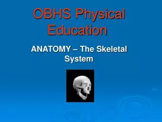
OBHS Physical Education ANATOMY – The Skeletal System
5 Major Functions There are five major functions of the skeleton: 1. Shape and support 2. Movement 3. Protection 4. Blood Production 5. Storage Remember: Should Mothers Protect Babies Skins
Shape and Support - This is our body's framework. It provides shape for our body, holds our vital organs in place and allows us to have a good posture. • Movement - Our muscles are attached to our bones in a way • which allows movement. • Protection - Protects our delicate organs e.g.-SKULL protects • the BRAIN. RIB CAGE protects the HEART and LUNGS etc. • Blood Production - Red and white blood cells are • produced in the bone marrow found in many bones. • RED CELLS carry oxygen to the muscles to enable them to • work. They are red in colour because they carry haemoglobin. • WHITE CELLS fight infection in the body. • Storage- minerals like calcium are stored in the bones to add strength
1. Cranium 2. Scapula 3. Clavicle 4. Humerus 5. Pelvis 6. Sternum 7. Ribs 8. Vertebrae 9. Radius 10. Ulna 11. Carpals & Metacarpals 12. Phalanges 13. Femur 14. Patella 15. Tibia 16. Fibula 17. Tarsals & Metatarsals 18. Phalanges Main Bones
Don’t be confused… The Chest Clavicle – collarbone Scapula – shoulder blade The Foot Metatarsals - foot Tarsals - ankle–think “T”for “toes” The Arm Radius - thumb side lower Ulna - finger side lower Humerus - upperarm – “funny bone” The Hand Carpals - wrist bones Metacarpals – hand The Leg Fibula - small lower Tibia - large lower Patella - knee
Joints Where bones meet they form JOINTS. The movement of the skeleton is helped by joints. There are THREE kinds of joints: • Fibrous (non-moving e.g.- skull) • Cartilagenous (limited movement e.g.-vertebrae of spine) • Synovial (a range of movements are available)
Synovial Joints Most moving joints are SYNOVIAL JOINTS. They are very complex structures. The Bones are linked together by ligaments and allow a wide range of movements. Features of a synovial joint include: Synovial fluid – Lubricates the joint Synovial Membrane – Seals the joint Synovial Capsule - Surround the joint to prevent leakage The knee is an example of a synovial joint
Connective tissue Joints are moved by muscles and bones. These are attached by LIGAMENTS and TENDONS. LIGAMENTS attach bone to bone. TENDONS attach muscle to bone. e.g.- The knee joint. Movements other than flexion/extension can cause serious ligament damage in hinge joints like the knee. In contact sports like rugby these ligaments are often strained by forces acting in other directions.
Joints Cont’d Joints can be separated into FOUR categories: Ball and Socket joint Hinge joint Gliding joint Pivot joint
Ball and Socket Two examples of this joint in the human body are the hip and shoulder joints. The rounded head of one bone fits into a cup-shaped socket of another. This joint allows the greatest range of movement. Pelvis Femur
Hinge Two examples of this type of joint include those found at the knee and elbow. Try flexing (bending) and extending them. You will find that the movement of the joint can only occur in one direction, just like the hinge of a door. Radius Humerus Ulna
Gliding In this type of joint, two surfaces which are flat rub against each other. These small bones can move over one another to increase flexibility of the hands for example. They are stopped from moving too far by strong ligaments. Carpals
Pivot This joint is made when one bone twists against another. These are found in the spine. They also allow the head to turn, raise and lower.
Types of Movement There are many types of movement that the skeleton and muscles can produce. The following are the most common: • Flexion • Extension • Rotation • Abduction • Adduction • Dorsiflexion • Plantarflexion
Types of Movement cont’d FLEXION – Bending the joint. E.g. Bending the knee or elbow. BALL and SOCKET and HINGE are the main joint types that can produce this movement.
Types of Movement cont’d EXTENSION of a joint is where the joint is straightened. BALL and SOCKET and HINGE joints are common examples of joints that can produce this movement. Straightening the leg when striking a ball is an example of EXTENSION at the knee (HINGE JOINT)
Types of Movement cont’d The ROTATION movement can occur at a BALL and SOCKET and a PIVOT joint. e.g. turning the head or the movement at the shoulder when swimming backstroke.
Types of Movement cont’d ABDUCTION and ADDUCTION movements can be produced by BALL and SOCKET joints. ABDUCTION is where a limb moves away from the centre of the body. ADDUCTION is where the limb is moved TOWARDS the centre of the body.
Joints and Performance Injuries to joints can occur from: • Over use (Too much training) • Incorrect movement injuries (e.g.-wrong techniques) • Impact or twisting (e.g.-twist of knee or elbow from a tackle or collision) Such injuries should be iced immediately, given plenty rest, elevated and compressed to aid recovery and avoid permanent damage. sports injuries clip