Download
1 / 66
680 likes | 1.05k Views
The biochemistry of cell injury and cell death. Dr Stephany Veuger. Overview. Part A Review causes of cellular damage Types of cellular damage Mechanisms of cell death Biochemical events that lead to cell death Part B Free radicals Diseases associated with free radical damage.

E N D
The biochemistry of cell injury and cell death Dr Stephany Veuger
Overview Part A • Review causes of cellular damage • Types of cellular damage • Mechanisms of cell death • Biochemical events that lead to cell death Part B • Free radicals • Diseases associated with free radical damage
Learning Outcomes • Understand how the basic functions of the cell are affected by injury • Discuss morphological and biochemical changes in response to injury • Be able to explain the types of cell death • Describe the biochemical changes in response to ischaemia
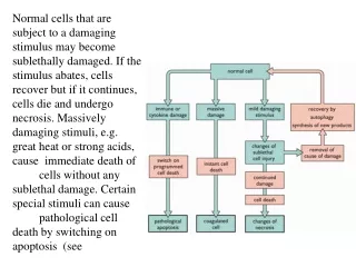
Causes of cell injury • Physical • Chemical • Infectious • Immunologic • Genetic derangement • Nutritional and Oxygen Imbalances • Metabolic changes
Cellular damage SUBLETHAL • Damage is minimal • Recovery LETHAL • Continued damage • Damage is massive
Mechanisms of cell injury Injurious agents can affect the cell at a number of levels by damaging : • Plasma membrane • Aerobic respiration and ATP production • Protein synthesis • Genetic machinery
Morphological indicators of cell injury • Alterations to plasma membrane • Cytoskeleton damage • Mitochondrial condensation • Mitochondrial swelling • Dilatation of ER • Ribosome detachment • Alterations to lysosomes
Morphological changes following sub-lethal injury • Mitochondrial swelling (low amplitude swelling) -vacuoles distort cristae -reversible • ER swelling -loss of ribosomes • High amplitude swelling -cristae destroyed -irreversible ATP-dependent processes affected
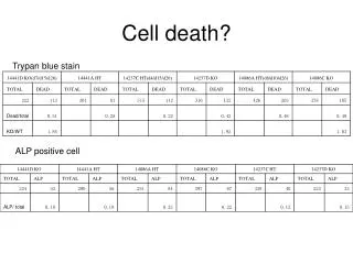
Morphological changes following sub-lethal injury Under the microscope, these changes are seen as; • Cellular swelling • Pale cytoplasm • Small intracellular vacuoles CLOUDY SWELLING or HYDROPIC DEGENERATION • Accumulation of lipid FATTY CHANGE
Fatty Change • Deficiency in lipid acceptor proteins, preventing export of formed triglycerides -carbon tetrachloride, malnutrition, hypoxis • Increased mobilisation of free FA into cells - diabetes mellitus and nutritional deprivation • Increased conversion of fatty acids to triglycerides -alcohol abuse • Reduced oxidation of triglycerides to acetyl-coA -hypoxia, toxins
Cell survival • Following injury, major cellular components need to be maintained to promote survival ; • Cell membranes • Mitochondria • Cytoskeleton • Cellular DNA - These systems are not interdependent - Threshold – death
Plasma membrane Integrity following injury is ESSENTIAL • Direct • Failure of phospholipid biosynthesis • Particularly vulnerable to free radical attack • Degradation of phospholipids by Ca2+ dependent phospholipases
Morphological changes following lethal injury • High amplitude swelling • Morphological changes to the nucleus • Appearance of membrane blebs and holes • Dissolution of the nucleus • Distinct structural changes to cell leading to dissolution of cell via release of lysosomal enzymes • AUTOLYSIS
Morphological changes following lethal injury (nucleus) • PYKNOSIS -condensation of nuclear chromatin • Loss of nucleolus • KARRYORRHEXIS -fragmentation of the nucleus • KARYOLYSIS -complete dissolution of nuclear material
Summary I • Cell have limited capacity to adapt to change • Mild injury can be accommodated by cells but is evident by biochemical and morphological changes • Sub-lethal –reversible • Injury that is sufficient to cause morpholgical changes to the nucleus is usually lethal • Dissolution of nuclear and cytoplasmic contents is caused by the release of lysosomal enzymes
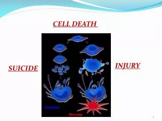
Cell death -Follows irreversible cell damage -Can be by accident or design • Apoptosis • Necrosis Different morphological changes
Apoptosis • Routine – repair and cell cycle (p53) • Programmed – co-ordinated- “shrinkage” • Stimuli mediated by immune system ; cytokines • Autophagy (self digestion)
Necrosis • Massive damage to cellular systems • Uncontrolled loss of large numbers of cells • Extensive organelle and cell “swelling” • Rupture of plasma membrane and dissolution of the cell
Biochemical determinants of necrotic change • ATP • Calcium homeostasis • pH • Reactive Oxygen Species (ROS) • Intracellular antioxidant levels
ATP • Produced by cellular respiration • biosynthesis • Critical for function of many transport pumps • Critical for cell signalling processes • Cloudy swelling and fatty change
Calcium • Normal concentration in cytosol very low -rapidly removed by ATP-dependent pumps -bound to buffering proteins (calbindin, parvalbumin) • Increased intracellular calcium brought about by; -↑permeability of Ca2+ channel -direct membrane damage -ATP depletion -mitochondrial damage
Cytosolic free calcium is a potent destructive agent CALCIUM STORES Mitochondria ER lumen Pumped to extracellular space Bound to binding proteins Released following cell injury FREE Ca 2+ Activation of ATPases Activation of phospholipases Activation of proteases Destabilising of cytoskeleton Reduced ATP Membrane damage
Reative Oxygen species (ROS) • Most important free radicals in the body are the oxygen-derived free radicals • Attack bio-molecules • Lipid peroxidation - decreases membrane fluidity and destabilises membrane receptors.
Effect of ROS on biomolecules GB.UNN.10
Changes in metabolism • Accumulation of materials as a result of changes in metabolism may compromise normal function of cell • Lipid (fatty change already covered) • Protein –kidneys, reversible • Carbohydrate-diabetes, glycogen storage disorders • pigments
ISCHAEMIA Excellent example of the cellular response to a damaging stimulus ISCHAEMIA = LACK OF OXYGEN SUPPLY HYPOXIA =LACK OF OXYGEN
Definitions • HYPOXIA -decrease in oxygen in arterial blood or tissues • ISCHAEMIA -local anaemia, leading to hypoxia eg. Obstruction to blood flow to organ/tissue • INFARCTION -sudden insufficiency of blood supply producing macroscopic areas of necrosis (eg. MI)
Biochemical and morphological changes due to Ischaemia (I) • Shift from aerobic to anaerobic respiration • Reduction in ATP • Failure of ATP-dependent pumps (Na+/K+, ATPase and Ca2+) • Failure to maintain intracellular ionic balance • Accumulation of Na+ in cytoplasm • Ingress of calcium and water and outflow of potassium ions Cloudy Swelling and disruption of internal membrane systems
Biochemical and morphological changes due to Ischaemia (II) Integrity of RER relies on Na+ pump • ribosomes detach • Protein synthesis ceases • Calcium – activation of several destructive enzyme systems • Phospholipid synthesis ceases Further disruption of membranes
Biochemical and morphological changes due to Ischaemia; pH (III) • Anaerobic respiration results in lactic acid production • Intracellular pH decreases • Membranes under acid attack • pH further augmented via phosphate ions produced by Ca2+ activated phosphatases • Fall in pH stimulates pyknosis
Biochemical and morphological changes due to Ischaemia; pH (IV) • Lysosomes • Release of destructive enzymes leads to karryhrrexis and karyolyiss • Cell death • Neighbouring cells injured • Initial changes in ischaemia reversible but nuclear changes catastrophic for cell
ISCHAEMIA Reduced oxidative phosphorylation Anaerobic respiration Decrease in sodium pump ?ATP Lactic acid ? ? pH ribosomes detach Potassium ? Calcium ?Protein synthesis ? ? lysosomes Water Cell death
ISCHAEMIA Reduced oxidative phosphorylation Anaerobic respiration Decrease in sodium pump ?ATP Lactic acid ? ? pH ribosomes detach Potassium ? Calcium ?Protein synthesis ? ? lysosomes Water Cell death
ISCHAEMIA Reduced oxidative phosphorylation Anaerobic respiration Decrease in sodium pump ATP Lactic acid ribosomes detach Potassium pH Calcium Protein synthesis pyknosis lysosomes Water karyorrhexis karyolysis Cell death
Summary II • Cells die by two main pathways • Biochemical determinants of injury and death ATP, Ca2+, pH, ROS • Ischaemia most common injury in clinical medicine
The role of free radicals and anti-oxidant mechanisms in health and disease
Overview • What are free radicals? • Sources of free radicals • Types of free radicals (ROS) • Types of free radical damage • Diseases associated with free radicals • Anti-oxidant mechanisms
Learning Outcomes • Define the terms free radical and reactive oxygen species • Characterise the major reactive oxygen species and their sources • Discuss the negative effects of ROS on bio-molecules • Describe the cellular defence mechanisms against free radicals
What is a free radical? • A radical is an atom or molecule with one or more unpaired electrons • A radical that can move freely within cell and across membranes is a free radical • Highly unstable and extremely reactive
Free radicals • Most molecules found in the body are not radicals. • Any reactive FR generated will often react with such non-radicals i.e. sugars, amino acids, phospholipids, nucleotides, polysaccharides, proteins, nucleic acids etc. • When this happens, a free radical chain reactionresults
Sources of free radicals • Ionising radiation • Chemicals • Exposure to excess oxygen • Cell respiration • Inflammation
Reactive oxygen species (ROS) GB.UNN.10
Abstraction Stripping of electrons from other atoms or molecules Propogation H abstraction on sugarssuch as deoxyribose yields many products, some of which are mutagenic. H abstraction on unsaturated membrane lipids is one of the most important aspects of damage to cells by FRs. R• + HB RH + B•
Addition • Attack of hydroxyl radical on DNA bases Thymine + OH● Thymine-OH● Hydroxythymine radical Thymine-OH● + OH● Thymine glycol
Effect on lipid • Peroxidation of membrane lipids is the most important cause of serious acute damage to cells Malondialdehyde = marker for oxidative stress • chain reaction of lipid peroxidation • H abstraction from a polyunsaturated fatty acid in a membrane or lipoprotein • Introduction of a polar group –OOH into hydrophobic region • Attack of one reactive FR can oxidise multiple fatty acid side chains to lipid peroxides
Effect on DNA • Reactive FRs such as the hydroxyl radical can react with both the deoxyribose and the bases of DNA • Thesugar component will be affected by H abstraction, resulting in many products, many of which are mutagenic. • Bases can be affected by addition reactions, ultimately leading to mutation and cellularderangement • Depletion of NADH pools
Effect on proteins • Formation of disulphide bridges by oxidation of the thiol groups (-SH) of cysteine residues • Attack metal binding sites leading to degradation by proteases • Loss of biological activity eg enzymes • Malondialdehyde - protein adducts or advanced lipoxidation end products (APE)