Download
1 / 19
190 likes | 348 Views
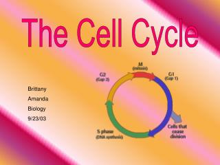
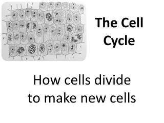
Biochemistry of the Cell Cycle . Topics Covered. What Drives the Cell Cycle? The Chemical Control of the Cycle Checkpoints Cyclin Controlled Cell Cycle Clock Loss of Control. What Drives the Cell Cycle?.

E N D
Topics Covered • What Drives the Cell Cycle? • The Chemical Control of the Cycle • Checkpoints • Cyclin Controlled Cell Cycle Clock • Loss of Control
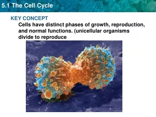
What Drives the Cell Cycle? • Hypothesis: The cell cycle is driven by specific molecular signals present in the cytoplasm. • Some of the first strong evidence for this hypothesis came from experiments with mammalian cells grown in culture. • In these experiments, two cells in different phases of the cell cycle were fused to form a single cell with two nuclei. • If one of the original cells was in the S phase and the other was in G1, the G1 nucleus immediately entered the S phase, as though stimulated by chemicals present in the cytoplasm of the first cell. • Similarly, if a cell undergoing mitosis (M phase) was fused with another cell in any stage of its cell cycle, even G1, the second nucleus immediately entered mitosis, with condensation of the chromatin and formation of a mitotic spindle
Chemical Control • There exists a cyclically operating set of molecules in the cell that both triggers and coordinates key events in the cell cycle. • The cell cycle control system has been compared to the control device of an automatic washing machine. • Like the washer′s timing device, the cell cycle control system proceeds on its own, driven by a built–in clock. • However, just as a washer′s cycle is subject to both internal control (such as the sensor that detects when the tub is filled with water) and external adjustment (such as activation of the start mechanism), the cell cycle is regulated at certain checkpoints by both internal and external controls.
Checkpoints • A checkpoint in the cell cycle is a critical control point where stop and go–ahead signals can regulate the cycle. (The signals are transmitted within the cell by the kinds of signal transduction pathways discussed in Chapter 11.) • Animal cells generally have built–in stop signals that halt the cell cycle at checkpoints until overridden by go–ahead signals. • Many signals registered at checkpoints come from cellular surveillance mechanisms inside the cell; the signals report whether crucial cellular processes up to that point have been completed correctly and thus whether or not the cell cycle should proceed. • Checkpoints also register signals from outside the cell. • Three major checkpoints are found in the G1, G2, and M phases.
Checkpoints • For many cells, the G1 checkpoint—dubbed the “restriction point” in mammalian cells—seems to be the most important. • If a cell receives a go–ahead signal at the G1 checkpoint, it will usually complete the S, G2, and M phases and divide. • Alternatively, if it does not receive a go–ahead signal at that point, it will exit the cycle, switching into a non-dividing state called the G0 phase . • Most cells of the human body are actually in the G0 phase. • Fully formed, mature nerve cells and muscle cells never divide. • Other cells, such as liver cells, can be “called back” from the G0 phase to the cell cycle by certain external cues, such as growth factors released during injury.
The Cell Cycle Clock • Rhythmic fluctuations in the abundance and activity of cell cycle control molecules pace the sequential events of the cell cycle. • These regulatory molecules are proteins of two main types: kinases and cyclins. • Protein kinases are enzymes that activate or inactivate other proteins by phosphorylating them . • Particular protein kinases give the go–ahead signals at the G1 and G2 checkpoints. • The kinases that drive the cell cycle are actually present at a constant concentration in the growing cell, but much of the time they are in an inactive form. • To be active, such a kinase must be attached to a cyclin, a protein that gets its name from its cyclically fluctuating concentration in the cell. • Because of this requirement, these kinases are called cyclin–dependent kinases, or Cdks . • The activity of a Cdk rises and falls with changes in the concentration of its cyclin partner. • The Cyclin-CDK complex is called the Maturation Promoting Factor (MPF). • Note that the peaks of MPF activity correspond to the peaks of cyclin concentration. The cyclin level rises during the S and G2 phases, then falls abruptly during mitosis (M).
How MPF Works • The initials MPF stand for “maturation–promoting factor,” but we can think of MPF as “M–phase–promoting factor” because it triggers the cell′s passage past the G2 checkpoint into M phase . • When cyclins that accumulate during G2 associate with Cdk molecules, the resulting MPF complex initiates mitosis, phosphorylating a variety of proteins. • MPF acts both directly as a kinase and indirectly by activating other kinases. • For example, MPF causes phosphorylation of various proteins of the nuclear lamina, which promotes fragmentation of the nuclear envelope during prometaphase of mitosis. • There is also evidence that MPF contributes to molecular events required for chromosome condensation and spindle formation during prophase. • During anaphase, MPF helps switch itself off by initiating a process that leads to the destruction of its own cyclin. • The noncyclin part of MPF, the Cdk, persists in the cell in inactive form until it associates with new cyclin molecules synthesized during the S and G2 phases of the next round of the cycle. • What about the G1 checkpoint? Recent research suggests the involvement of at least three Cdk proteins and several different cyclins at this checkpoint. The fluctuating activities of different cyclin–Cdk complexes seem to control all the stages of the cell cycle.
Signals to Grow and Divide • In many cases, though, scientists don′t yet know what the various Cdks actually do. However, they have identified some steps of the signaling pathways that convey information to the cell cycle machinery.
Signals to Grow and Divide--Internal • An example of an internal signal occurs at the M phase checkpoint. Anaphase, the separation of sister chromatids, does not begin until all the chromosomes are properly attached to the spindle at the metaphase plate. • Researchers have learned that kinetochores not yet attached to spindle microtubules send a molecular signal that causes the sister chromatids to remain together, delaying anaphase. • Only when the kinetochores of all the chromosomes are attached to the spindle will the sister chromatids separate (owing to inactivation of the proteins holding them together). This mechanism ensures that daughter cells do not end up with missing or extra chromosomes.
Signals to Grow and Divide--External • By growing animal cells in culture, researchers have been able to identify many external factors, both chemical and physical, that can influence cell division. • For example, cells fail to divide if an essential nutrient is left out of the culture medium. • And even if all other conditions are favorable, most types of mammalian cells divide in culture only if the growth medium includes specific growth factors. • Recall that a growth factor is a protein released by certain cells that stimulates other cells to divide. • While called a growth factor for historical reasons, a protein that promotes mitosis is sometimes more narrowly called a mitogen. • One such growth factor is platelet–derived growth factor (PDGF), which is made by blood cells called platelets.
PDGF Example • This example demonstrates that PDGF is required for the division of fibroblasts in culture. • Fibroblasts, a type of connective tissue cell, have PDGF receptors on their plasma membranes. • The binding of PDGF molecules to these receptors (which are receptor tyrosine kinases) triggers a signal transduction pathway that allows the cells to pass the G1 checkpoint and divide. • PDGF stimulates fibroblast division not only in the artificial conditions of cell culture, but in an animal′s body as well. • When an injury occurs, platelets release PDGF in the vicinity. The resulting proliferation of fibroblasts helps heal the wound. • Researchers have discovered at least 50 different growth factors that can trigger cells to divide. • Different cell types respond specifically to a certain growth factor or combination of growth factors.
Growth Inhibition • The effect of an external physical factor on cell division is clearly seen in density–dependent inhibition, a phenomenon in which crowded cells stop dividing .
Growth Inhibition • Cultured cells normally divide until they form a single layer of cells on the inner surface of the culture container, at which point the cells stop dividing. • If some cells are removed, those bordering the open space begin dividing again and continue until the vacancy is filled. • It was originally thought that a cell′s physical contact with neighboring cells signaled it to stop dividing. • However, while physical contact may have some influence, it turns out that the amount of required growth factors and nutrients available to each cell has a more important effect: Apparently, when a cell population reaches a certain density, the availability of nutrients becomes insufficient to allow continued cell growth and division. • Most animal cells also exhibit anchorage dependence. To divide, they must be attached to a substratum, such as the inside of a culture jar or the extracellular matrix of a tissue. • Experiments suggest that anchorage is signaled to the cell cycle control system via pathways involving plasma membrane proteins and elements of the cytoskeleton linked to them. • Density–dependent inhibition and anchorage dependence appear to function in the body′s tissues as well as in cell culture, checking the growth of cells at some optimal density and location. Cancer cells exhibit neither density–dependent inhibition nor anchorage dependence .