Muscle Contraction Biology
Explore the intricate details of muscle contraction, from sarcomere structure to neuromuscular junction function, in this comprehensive warm-up. Learn about the sliding filament model, muscle types, and cellular anatomy that drive movement.

Muscle Contraction Biology
E N D
Presentation Transcript
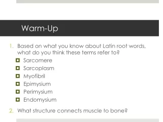
Warm-Up • Based on what you know about Latin root words, what do you think these terms refer to? • Sarcomere • Sarcoplasm • Myofibril • Epimysium • Perimysium • Endomysium • What structure connects muscle to bone?
Warm-Up • What is the organization of a skeletal muscle from the largest to the smallest structures? • Draw and label the parts of a sarcomere. Be sure to include the thick & thin filaments, I band, A band, and Z lines.
Warm-Up • Describe what happens at the neuromuscular junction. • How would a drug that blocks acetylcholine (ACh) release affect muscle contraction? • Which of the following pictures below shows a contracted muscle? Explain your answer.
Warm-Up Put the following events in muscle contraction in order: • Calcium binds to troponin changes shape myosin binding sites exposed on actin • Myosin head pivots and pulls actin filament toward M line • ATP attaches to myosin and cross-bridge detaches • Action potential travels down sarcolemma along T-Tubules • Myosin cross-bridge forms with actin • Calcium is released from sarcoplasmic reticulum (SR)
Warm-Up • Jay is competing in a chin-up competition. What types of muscle contractions are occurring in his biceps muscles: • immediately after he grabs the bar? • as his body begins to move upward toward the bar? • when his body begins to approach the mat? • When a suicide victim was found, the coroner was unable to remove the drug vial from his hand. Explain.
Muscles & Muscle Tissue Chapter 9
Muscles • “muscle” = myo- or mys- • sarco- = “flesh” - also refers to muscles
Main Functions of Muscles • Produce movement • Maintain posture & body position • Stabilize joints • Generate heat Additional: protect organs, valves, dilate pupils, raise hairs
Types of Muscle Tissue • Skeletal: voluntary, striated, multinucleated • Cardiac: (heart) striated, involuntary • Smooth: visceral (lines hollow organs), nonstriated, involuntary
Special Characteristics • Excitability – can receive and respond to stimuli • Contractility – can shorten forcibly • Extensibility – can be stretched or extended • Elasticity – can recoil and resume resting length after being stretched
Gross Anatomy of Skeletal Muscle • 1 muscle = 1 organ • Each muscle served by a nerve, artery, & vein (1+) • Rich blood supply– need energy & O2 • Connective tissue sheaths: wraps each cell and reinforce whole muscle • Attachment: (1) directly to bone, (2) by tendons or aponeuroses to bone, cartilage, or other muscles
Muscle • Muscle cells + blood vessels + nerve fibers • Covered by epimysium (connective tissue) Organization of Skeletal Muscle • Fascicle • Bundle of muscle cells • Surrounded by perimysium • Muscle fiber (cell) • Surrounded by endomysium • Myofibril • Complex organelle • Sarcomere • Contractile unit
Gross Anatomy of Skeletal Muscle Video Clip
Anatomy of Muscle Fiber • Multinucleate cell • Up to 30 cm long • Sarcolemma (plasma membrane) • Sarcoplasm (cytoplasm) • Myofibril = rodlike organelle • Contains contractile element (sarcomeres) • Alternating light (I)and dark (A)bands
Sarcomere • Smallest contractile unit of muscle fiber • Region between 2 successive Z discs
Sarcomere • Protein myofilaments: • Thick filaments = myosin protein • Thin filaments = actinprotein
Myofilaments Thick Filaments Thin Filaments Tropomyosin: protein strand stabilizes actin Troponin:bound to actin, affected by Ca2+ • Myosin head: forms cross bridges with thin filaments to contract muscle cell
Sarcoplasmic Reticulum (SR): specialized smooth ER, surrounds each myofibril • Stores and releases calcium • T Tubule: part of sarcolemma, conducts nerve impulses to every sarcomere • Triggers release of calcium from SR
Sliding Filament Model • During contractions: thin filaments slide past thick ones so they overlap more
Sliding Filament Model • Myosin heads latch onto active sites on actin to form a cross-bridge • Attachments made/broken tiny rachets to propel thin filaments to center of sarcomere
Skeletal Muscle Structure Video Clip
Basic Muscle Contraction • Stimulation by nerve impulse • Generate and send electrical current (action potential) along sarcolemma • Rise in calcium ion levels to trigger contraction
Nerve Impulse • 1 nerve cell (motor neuron) stimulates a few or hundreds of muscle cells • Motor unit = 1 neuron + muscle cells stimulated • Axon: extension of neuron • Axon terminal: end of axon • Neuromuscular junction (NMJ): where axon terminal meets muscle fiber • Synpatic cleft: space between neuron & muscle fiber • Acetylcholine (ACh): neurotransmitter
Excitation of Muscle Cell • Action potential travels down axon and arrives at neuromuscular junction • Release of acetylcholine (ACh) into synaptic cleft • ACh diffuses across cleft & attaches to ACh receptors on sarcolemma of muscle fiber • Rush of sodium (Na+) into sarcoplasm produces action potential in sarcolemma • ACh broken down
Contraction of Muscle Cell • Action potential travels down sarcolemma along T-Tubules • Calcium is released from SR • Calcium binds to troponin changes shape myosin binding sites exposed on actin • Myosin cross-bridge forms with actin • Myosin head pivots and pulls actin filament toward M line • ATP attaches to myosin and cross-bridge detaches • Myosin can be reactivated
Action Potentials and Muscle Contraction Video Clip
Neuromuscular Junction Video Clip
Homeostatic Imbalances • Myasthenia gravis: loss of ACh receptors in sarcolemma by immune system attack progressive muscular paralysis • Botulism: from bacterial toxin; prevents release of ACh at synaptic terminals muscular paralysis • Rigor mortis: “death stiffness” = no ATP production, myosin cross-bridges “stuck” until proteins break down (peak: 12 hrs, fades: 48-60 hrs later)
Muscle Responses • Twitches(single, brief, jerky contractions) = problem • Healthy muscle = smooth contraction • Graded muscle responses: different degrees of muscle shortening Greater force by: • Increase frequencyof muscle stimulation • Contractions are summed (max tension = complete tetanus) • Increase # muscle cells being stimulated
Energy • ATP= only energy source for muscles • Regenerated by: • Creatine phosphate (CP): transfers energy to ATP • Aerobic respiration: complete glucose breakdown with O2 present • Lactic acid fermentation: glucose breakdown without O2 • Muscle fatigue: lack of O2, ATP supply low, lactic acid accumulates, soreness muscle contracts more weakly until it stops
Creatine Phosphate Supplements • Muscle cells store phosphocreatine (Pcr) for sprinting and explosive exercise • Forms/uses: powders, tablets, energy bars, drink mixes • Supplements can enhance sprint performance and lean muscle mass (no evidence to aid endurance performance) • Side effects: weight gain, anxiety, diarrhea, fatigue, headache, kidney problems, nausea, vomiting, rash • No recommended for peoplewith diabetes, kidney or liver problems • Caution: Drink lots of water to avoid dehydration • Effects of long-term usage: unknown
Types of Contractions Isotonic Isometric “same length” Muscle length stays same Tension increases Moving against heavy load or immovable object Eg. lifting heavy weights • “same tension” • Muscle length changes • Concentric: shortens • Eccentric: lengthens • Eg. bicep curl, bend knee, smiling
Muscle Tone • Muscles: firm, healthy, ready for action • Some fibers contracting even when muscle is relaxed Nerve Damage (paralysis): • Flaccid: muscles soft & flabby • Atrophy: wasting away if not stimulated
Exercise = Use it or lose it! • Aerobic (endurance) Exercise • stronger, more flexible muscles, greater resistance to fatigue • No increase in muscle size • blood supply, mitochondria, O2 storage • efficiency of metabolism, heart function • Eg. aerobics, jogging, biking
Exercise = Use it or lose it! • Resistance/Isometric Exercise • Muscles vs. immovable object • muscle cell size (more contractile filaments) • muscle size and strength • Eg. weights, using own body
Muscle Cramps • Sudden or involuntary contraction of muscles • Causes: long periods of exercise or physical labor, medications, dehydration, muscle strain, nerve/kidney/thyroid disorders • Medical Condition: Inadequate blood supply, nerve compression, mineral depletion (Ca, K, Mg) • Treatment: stretching exercises, muscle relaxant, hydration, Vitamin B supplements, apply cold/heat