Download
1 / 12
130 likes | 290 Views
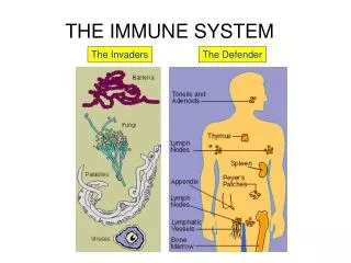
Blood and the Immune System. The Body’s Lines of Defence. First Line of Defence. Skin – the largest organ, and mucous membranes defend against viral and bacterial invaders.

E N D
Blood and the Immune System The Body’s Lines of Defence
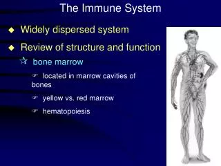
First Line of Defence Skin – the largest organ, and mucous membranes defend against viral and bacterial invaders. The skin acts as a physical barrier as wells as a chemical barrier – acidic secretions which inhibits the growth of microbes Lysozyme – antimicrobial enzyme secreted in tears, saliva, mucous secretions and perspiration destroys cell walls of bacteria Mucus in the respiratory tract trap foreign debris and invading microbes Cilia sweep particles up where coughing can expel them Corrosive acids in stomach and protein-digesting enzymes destroy most of invading microbes in food.
Second Line of Defence Leukocytes (WBC) engulf invading microbes Rely on process of phagocytosis. Monocytes migrate from blood to tissue and develop into macrophages. Extend pseudopods to attach to surface of microbe which is then engulfed and destroyed Neutrophils are attracted to chemical signals – chemotaxis is the process where neutrophils squeeze out of capillaries and migrate towards infected tissue. Engulf microbe and release lysosomalenzymes where both microbe and leukocyte are destroyed.
Pus and Inflammation Remaining fragments of protein, dead WBC and digested invaders after phagocystosis Non-specific response is seen as inflammation around the area where tissue has been damaged = swelling, redness, heat and pain
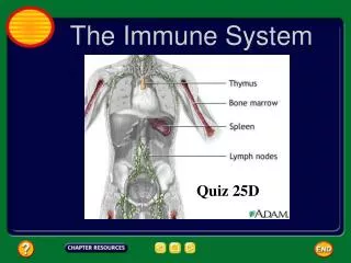
The Immune Response – Third Line of Defence Found localized in the brain, lungs, kidneys, liver and connective tissue Complement Proteins – antimicrobial plasma protein triggered by foreign bodies Act in three ways: Envelop and seal invader Attaches and punctures the cell membrane causing cell to swell and burst - Attaches to invader attracting leukocytes
Lymphocytes WBC that produce antibodies Foreign bodies contain many antigens on their surface. T-cell lymphocytes are produced in bone marrow stored in thymus gland Seeks out intruder and signals attack B-cell leukocytes are anti-body producing. Each B-cell produces a single type of antibody. Super-antibody-producing cells are called plasma cells which produce 2000 antibody molecules/sec
Antigen-Antibody Reactions Y-shaped proteins that are specific to the foreign invaders. Antibodies produced against influenza will not protect against HIV Antibodies only attach to its complementary marker creating a larger complex making it an easier target for macrophages to engulf and destroy Toxins are prevented from prevented from destroying cells when the antibody binds to the toxin and interferes with the attachment of toxins to the cell.
Viruses and Mutations Use receptor sites as points of entry Injects hereditary material into cell but leaves an outer protein coat at receptor site. HIV attaches to receptor sites of T-cells. The virus is engulfed but since the blue-print for the antibody has been engulfed, it is not recognized as being a foreign invader. Antibody may cause virus to change shape. May only change slightly because of a mutation which allows the virus to still gain access to receptor site without being recognized by an antibody
Recognizing Harmful Antigens Foreign antigen markers are not destroyed in engulfment Pushed to cell membrane of macrophage where the antigen is couples with helper T cells Lymphokine – chemical messenger released by T cells reading antigen’s shape B cells are encouraged to divide based on lymphokine Second message is released by helper T cells to B cells triggering production of antibodies Helper T cells trigger killer T cells = search and destroy missions They go after viruses by detecting the viral coat and attacking the infected cell.
Killer T Cells Destroy mutated cells – attack pre-cancerous cells Antigen markers on cell membrane of donor tissue different from recipient – an assault is issued. Immunosuppressant drugs (cyclosporin) are given to slow down Killer T cells -will become susceptible to bacterial infections Suppressor T Cells inhibits immune response Helper T cells and Suppressor T cells spend a lot of time on the ‘cell’ phone talking to each other
Immune System Memory 18-19th Century Aboriginal populations were greatly impacted after coming into contact with European settlers carrying the small pox virus No antibodies present Helper T cells reads a blueprint of invader before B cells produce antibodies Memory B cell is generated during infection – holds an imprint of antigen T and B cells die off days after fight, but memory B cells stick around and can identify invader at a later date.