Download
1 / 36
400 likes | 2.14k Views
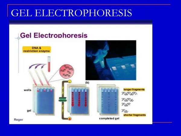
Electrophoresis and 2D Gel Analysis. What is Electrophoresis ? The migration of a charged particle (protein) through a separation matrix toward the opposite charged electrode. Anion. Cation. Anode. Cathode. Types of Electrophoresis. 1D Gel Electrophoresis- DNA, RNA, and Protein

E N D
What is Electrophoresis ?The migration of a charged particle (protein) through a separation matrix toward the opposite charged electrode Anion Cation Anode Cathode
Types of Electrophoresis 1D Gel Electrophoresis- DNA, RNA, and Protein Agarose –low resolution, general use for RNA and DNA Polyacrylamide – High resolution Size determination, DNA Sequencing, gel shift, etc SDS-PAGE-Protein Pulse Field- DNA separation of very large fragments of genomic DNA 2D Gel Electrophoresis Isoelectric focus and PAGE –Protein DIGE-Differential gel electrophoresis Two samples labeled with different fluorophores are mixed and run on a single 2d gel Capillary Electrophoresis (CE) Linearized polyacrylamide, PVP Automated DNA Sequencing, genotyping Micro-channel/ Micro-fluidic Electrophoresis Lab-on-a-Chip
(-) (-) (-) (-) (-) slower migration faster migration Electrophoresis and Biomolecules DNA and RNA: Phosphate backbone is negatively charged (anionic) and moves toward positive charge (anode) Separated based on size and sometimes structure
Electrophoresis and Biomolecules Proteins: Proteins can have both positive and negative charges Proteins can be acidics or basic [amphoteric] In their native state, the overall charge is variable Anionic detergents [SDS] denature secondary and non–disulfide–linked tertiary structures and impart a negative charge in proportion to its mass. SDS is therefore required when running standard gels to determine size in KDa
Electrophoresis and Biomolecules Proteins: Isoelectric Point (IEP)- also known as pI pH at which a protein has a neutral charge loss or gain of protons H+ in a pH gradient That is to say… In a pH below their pI, proteins carry a net positive charge and for in a pH above their pI, they carry a net negative charge.
Proteins: Isoelectric Focusing (IEF): “First Dimension” in 2-D gel Utilizes a special pH gradient strip and high voltage electrophoretic equipment Variable pH Ampholytes embedded in Acrylamide Proteins separate of according to their isoelectric point (pI) The equilibrium that a protein establishes at a specific pH on the IPG is an electro-chemical “focusing”. When a protein moves away from its IEF pH, its charge will deviate from a neutral state and either gain charge or lose charge and finding its way back to the “comfortable” state of neutral. IEF apparatus from Bio-Rad
1D Gel Electrophoresis-SDS PAGEStandard use for protein separation based on size (MW)Uses sodium dodecyl sulfate to denature protein and provide negative charge for mobility Without SDS, different proteins with similar molecular weights would migrate differently due to differences in mass charge ratio and structure Degree of resolution determined by % acrylamide
_ + SO Na 4 Sodium Dodecyl Sulfate _ _ _ _ _ _ _ C A T H O D E A N O D E + + + + + + + _ _ _ _ _ _ _ _ _ _ _ _ _ _ _ _ _ _ _ _ _ _ _ _ _ _ _ _ _ SDS PAGE Principles
Components of SDS PAGE Gel B-Mercaptoethanol /DTT [a reducer] Added to prevent oxidation of cysteines and to break up disulfide bonds within the proteins Sodium dodecyl sulfate [negatively-charged ionic detergent] binds to the vast majority of proteins at a constant ratio of 1.4 gm SDS/gm protein and provide a constant charge to mass ratio for electro-mobility Glycerol Provides density for gel loading Bromophenyl blue A tracking dye used for visualization purposes during a run
Components of SDS PAGE Gel Stacking Gel Is prepared w/Tris/HCL buffer pH 6.8, ~2pH units lower than running buffer. Large pore polyacrylamideused to align and create a thin starting zone of the protein of apx. 19um on top of the resolving gel. Resolving Gel Small pore polyacrylamide gel (3 - 30% acrylamide monomer) typically made using a pH 8.8 Tris/HCl buffer. Resolves protein ~24 – 205 kDa Running Buffer Tris/Glycine: Glycine(pKa=9.69) is a trailing ion (or slow ion). In other words it runs through the gel slower then the slowest protein at a pH above 8.0.
Polyacrylamide Chemistry Acrylamide monomer Tetramethylethylene diamine [TEMED] Catalyst {O2 Scavenger} N,N'-methylene-bis-acrylamide Ammonium persulfate {Free radical generator} Polyacrylamide TEM image Pore size of gel is determined by total amount of acrylamide and bis-acrylamide
200 200 200 45 200 45 45 200 45 6.5 45 Affect of % Acrylamide
Staining Polyacrylamide Gels Coomassie Blue Stain-can usually detect a 10-50 ng protein per band Blue Safe Stains -Similar to Coomassie. Destaining optional or water rinse. (8 ng/band) BioSafe Blue SimplyBlue GelCode InstantBlue-destaining not recommended Silver Staining-50 times more sensitive than Coomassie Blue. (0.3ng/BAND) Fixation [Acetic acid-methanol] Sensitize gel with sodium thiosulfate Stain with silver solution Rinse with water Develop with formaldehyde and carbonate followed by stopping with Glacial acetic Fluoresecnt Stains –almost as sensitive as Silver but requires excitation source Flamingo Fluorescent Gel Stain Deep Purple* Total Protein Stain SYPRO* Ruby Protein Gel Stain Krypton Protein Stain IR stains
Coomassie –Protein Binding Sulfonic acid group interacts with positively charged amine R groups Basic amino acids including arginine, lysine and histidine but weakly with histidine, tyrosine, tryptophan and phenylalanine Interactions in its anionic form [-] Electrostatic Ionic Van der Waals Hydrophobic
Destaining Gels Most gels require destaining to see banding and to eliminate background stain for high resolution. Gels with abundant protein need not be destained when using certain SafeBlue stains such as InstantBlue Coomassie blue destaining Usually requires acetic acid , methanol, and water Safe Blue destaining Usually requires water rinse Silver Stains Some methods use Potassium Ferricynide -Sodium Thiosulfate solutions Some methods use Sodium chloride -Cupric sulfate -Sodium thiosulfate pentahydrate. Some destaining may require a stop solution including 10% Acetic acid
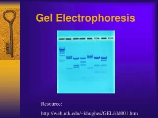
2D Gel Electrophoresis Yeast Proteome: 50 ug protein loaded, pH 4-8 ampholines, 10% slab gel, silver stain.
2D Gel Electrophoresis Separation of hundreds of proteins based on -pI -MW Up to 10,000 proteins can be seen using optimized protocols
Why 2D Gels Oldest method for large scale protein separation (since 1975) Popular method for protein display and proteomics-one spot at a time Can be used in conjunction with Mass Spec Permits simultaneous detection, display, purification, identification, quantification, pI, and MW. Robust, reproducible, simple, cost effective, scalable Provides differential quantification using Differential 2D Gel Electrophoresis (DIGE)
Processes involved in 2D gel electrophoresis Protein isolation and quantification Isoelectric focusing (first dimension) SDS-PAGE (second dimension) Visualization of proteins spots with Dye Identification of protein spots with Mass Spec Bioinformatics
Sample Preparation • Sample preparation is key to successful 2D gel experiments • Must select appropriate method to get selected proteins from cellular compartment of interest • Membrane proteins, nuclear proteins, and mitochodrial proteins require special steps • Must break all non-covalent protein-protein, protein-DNA, protein-lipid interactions, disrupt S-S bonds • Must prevent proteolysis, accidental phosphorylation, oxidation, cleavage, ect.. • Must remove substances that might interfere with separation process such as salts, polar detergents (SDS), lipids, polysaccharides, nucleic acids • Must try to keep proteins soluble during both phases of electrophoresis process • Must quantify protein
Protein Solubilization 2-20 mM Tris base (Carrier ampholytic buffer) 5-20 mM DTT (to reduce disulfide bonds) 8 M Urea (neutral chaotrope) Increases the solubility of some proteins Chaotropic agents interfere with stabilizing non-covalent forces (hydrogen bonds, van der Waals forces, and hydrophobic) 4% CHAPS Detergent(3-[(3-Cholamidopropyl)dimethylammonio]-1-propanesulfonate) pH of 5-7 Zwitterionic detergent (electronically neutral-has a both Neg and Pos useful for varible charged peptides ) Protects the native state of proteins Better when downstream apps include IEF because no affect on pH gradients
pH 3 4 5 6 7 8 9 10 IEF and IPG (immobilized pH Gradient) Strip of paper Made by covalently integrating acrylamide and variable pH ampholytes Separation on basis of pI, not MW Available in different pH ranges 3-10 4-8 5-7 Requires very high voltages (5000V)and long period of time (10h)
I IPG Strips Contain Ampholytes Ampholytes are molecules that contain both acidic and basic groups Protein will migrate in the Matrix and will find their pH equilibrium (pI)
SDS Gel Negative electrode IPG strip-pressed down into the SDS-PAGE gel pH3 4 5 6 7 8 9 10 Similar pI but different mw Similar mw but different pI Positive electrode
Different IPG pH ranges yield Different Results pH 4 pH 5 pH 4 pH 9 pH 5 pH 7
Gel Stains - Summary Stain Sensitivity (ng/spot) Advantages Coomassie-type 5-10 Simple, fast Silver stain 1-4 Very sensitive, laborious Copper stain 5-15 Reversible, 1 reagent negative stain Zinc stain 5-15 Reversible, simple, fast high contrast neg. stain SYPRO ruby 1-10 Very sensitive, fluorescent
2D Gel Results • 401 spots (peptides or PTM) identified • 279 gene products
2D Gel Post Analysis Compare gel images and determine what bands/spots are different Requires software to compare gels Apparent difference- Need to extract spot for MS
Extracting a Gel Spot Run Mass spec Cut out spot Trypsin Digestion of Gel spot
Differential 2D Gel Electrophoresis [DIGE]Allows you to mix samples and run a single 2d gel for comparative and quantitative purposes Fluorescent stain Cy3--Normal liver Cy5--Tumor Both
Conclusions • 2D gel electrophoresis is a popular method for protein display, separation, visualization, and quantitation • A good precursor to MS, but not required • 2D gels provide pI, MW data, and photodocumentation • Web tools are now available that permit partial analysis and comparison of 2D gels using software and simulators • 2D gels are fun to run