Gel Electrophoresis
340 likes | 727 Views
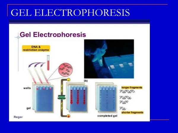
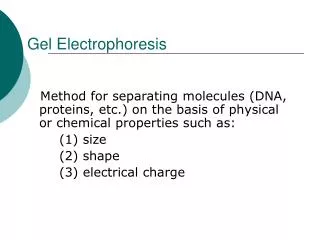
Gel Electrophoresis. The questions. Why? To separate pieces of DNA by size. How? Use electrical charge to pull the negatively charged DNA through a gel that has small pores in it.

Gel Electrophoresis
E N D
Presentation Transcript
The questions • Why? To separate pieces of DNA by size. • How? Use electrical charge to pull the negatively charged DNA through a gel that has small pores in it. • When? When doing DNA fingerprinting, analysis of plasmids, determining sizes of fragments, mapping plasmids
• When placed in an electrical field, DNA will migrate toward the positive pole (anode). H á O2 á DNAà - + Power • Polymerized agarose is porous, allowing for the movement of DNA Scanning Electron Micrograph of Agarose Gel (1×1 µm) à • DNA is negatively charged. • An agarose gel is used to slow the movement of DNA and separate by size.
DNA - + Power How fast will the DNA migrate? strength of the electrical field, buffer, density of agarose gel… Size of the DNA! *Small DNA move faster than large DNA …gel electrophoresis separates DNA according to size small large Within an agarose gel, linear DNA migrate inversely proportional to the log10 of their molecular weight.
Buffer Buffer is to keep the pH constant. If the pH changed the H+ charges would be pulled to the negative cathode and would set up a pH gradient in the box.
Agarose D-galactose 3,6-anhydro L-galactose • Sweetened agarose gels have been eaten in the Far East since the 17th century. • Agarose was first used in biology when Robert Koch* used it as a culture medium for Tuberculosis bacteria in 1882 *Lina Hesse, technician and illustrator for a colleague of Koch was the first to suggest agar for use in culturing bacteria Agarose is a linear polymer extracted from seaweed.
Agarose vs Agar • Agar: from seaweed is food for bacteria to grow upon • Agarose: sugar gel for doing electrophoresis
An agarose gel is prepared by combining agarose powder and a buffer solution. BufferØ Flask for boilingÙ AgaroseÚ
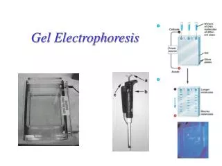
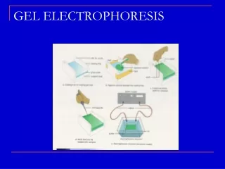
Electrophoresis Equipment Power supplyà åCover Gel tankà Electrical leads â Casting trayä Gel combsä
Preparing the Casting Tray Seal the edges of the casting tray and put in the combs. Place the casting tray on a level surface. None of the gel combs should be touching the surface of the casting tray.
Agarose Buffer Solution Combine the agarose powder and buffer solution. Use a flask that is several times larger than the volume of buffer.
Melting the Agarose Agarose is insoluble at room temperature (left). The agarose solution is boiled until clear (right). Gently swirl the solution periodically when heating to allow all the grains of agarose to dissolve. ***Be careful when boiling - the agarose solution may become superheated and may boil violently if it has been heated too long in a microwave oven.
How thick? • Agarose is measured in %, which is grams per 100ml of solution. • A 7% gel vs a 3 % gel; • Thicker so it is easier to hold without breaking • Slower for molecules to go through and hard for big molecules to make it.
Pouring the gel Allow the agarose solution to cool slightly (~60ºC) and then carefully pour the melted agarose solution into the casting tray. Avoid air bubbles.
Each of the gel combs should be submerged in the melted agarose solution.
When cooled, the agarose polymerizes, forming a flexible gel. It should appear lighter in color when completely cooled (30-45 minutes). Carefully remove the combs and tape.
DNAè bufferè á á á á wells Anodeä (positive) ãCathode (negative) Add enough electrophoresis buffer to cover the gel to a depth of at least 1 mm. Make sure each well is filled with buffer.
Sample Preparation Mix the samples of DNA with the 6X sample loading buffer (w/ tracking dye). This allows the samples to be seen when loading onto the gel, and increases the density of the samples, as it has glycerol added to increase the density to greater than water. This causes the dye and DNA sample to sink into the gel wells. Remember the wells are like little cups, open at the top and walls on each side. 6X Loading Buffer: à · Bromophenol Blue (for color) · Glycerol (for weight)
Loading the Gel Carefully place the pipette tip over a well and gently expel the sample. The sample should sink into the well. Be careful not to puncture the gel with the pipette tip.
Running the Gel Place the cover on the electrophoresis chamber, connecting the electrical leads. Connect the electrical leads to the power supply. Be sure the leads are attached correctly - DNA migrates toward the anode (red). When the power is turned on, bubbles should form on the electrodes in the electrophoresis chamber.
Cathode (-) ß wells ß Bromophenol Blue DNA (-) â Gel Anode (+) After the current is applied, make sure the Gel is running in the correct direction. Bromophenol blue will run in the same direction as the DNA.
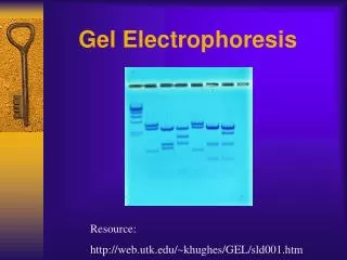
ß 12,000 bp ß 5,000 DNA migration ß 2,000 ß 1,650 ß 1,000 ß 850 ß 650 ß 500 ß 400 ß 300 ß 200 ß 100 DNA Ladder Standard - Note: bromophenol blue migrates at approximately the same rate as a 300 bp DNA molecule bromophenol blueà + Inclusion of a DNA ladder (DNAs of know sizes) on the gel makes it easy to determine the sizes of unknown DNAs.
Staining the Gel • Ethidium bromide binds to DNA and fluoresces under UV light, allowing the visualization of DNA on a Gel. • Ethidium bromide can be added to the gel and/or running buffer before the gel is run or the gel can be stained after it has run. ***CAUTION! Ethidium bromide is a powerful mutagen and is moderately toxic. Gloves should be worn at all times.
Safer alternatives to Ethidium Bromide • · Methylene Blue • · BioRAD - Bio-Safe DNA Stain • Ward’s - QUIKView DNA Stain • Carolina BLU Stain • …others advantages Inexpensive Less toxic No UV light required No hazardous waste disposal disadvantages Less sensitive More DNA needed on gel Longer staining/destaining time
Staining the Gel • Place the gel in the staining tray containing warm diluted stain. • Allow the gel to stain for 25-30 minutes. • To remove excess stain, allow the gel to destain in water. • Replace water several times for efficient destain.
Ethidium Bromide requires an ultraviolet light source to visualize
DNA ladder â DNA ladder â 1 2 3 4 5 6 7 8 wellsà • 5,000 bp ß 2,000 ß 1,650 ß 1,000 ß 850 ß 650 ß 500 ß 400 PCR Product ß 300 ß 200 ß 100 + - - + - + + - Visualizing the DNA (ethidium bromide) Primer dimersà Samples # 1, 4, 6 & 7 were positive for Wolbachia DNA
Visualizing the DNA (QuikVIEW stain) DNA ladder â wellsà ß 2,000 bp PCR Product ß 1,500 ß 1,000 ß 750 ß 500 ß 250 + - - - - + + - - + - + Samples # 1, 6, 7, 10 & 12 were positive for Wolbachia DNA March 12, 2006
Problem? • Ethidium Bromide is a carcinogen • Radioactivity is a carcinogen and is hard to get • Blue stains smear and are much harder to see. • In college: Bio 101 would use blue • Molecular Biology: would use Ethidium bromide.