Platelet Physiology and Disorders: Understanding Structure, Function, and Diseases
This comprehensive guide delves into platelet physiology, production, circulation, adhesion, and disorders such as thrombocytopenia and immunothrombocytopenia. Explore platelet structure, mechanisms, and diseases related to bone marrow suppression, DIC, and aplastic anemia. Gain insights into autoimmune platelet disorders, manifestations, and laboratory findings.


Platelet Physiology and Disorders: Understanding Structure, Function, and Diseases
E N D
Presentation Transcript
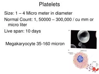
PLETELET PHYSIOLOGY Platelets Production: Hematopoietic stem cell Megakaryoblast Megakaryocyte Fragmentation of cytoplasm Platelets
Thrombopoietin: • Regulator of platelet production. • Produced by the liver and kidneys. • Levels are increased in thrombocytopenia,and reduced in thrombocytosis. • It increases the no. & rate of maturation of the megakaryocytes.
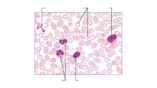
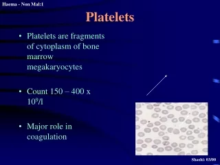
PLATELET CIRCULATION • Normal count is 250,000. • Normal life span 7-10 days. • About 1/3 are trapped in the spleen.
STRUTURE Mucopolysacch. coat Granules Dense core granules Mucopolysacch. Coat: Glycoprotein content which are important for interaction of platelets with each other or aggregating agents. • Granules: • Dense core:
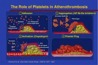
Function Formation of mechanical plug during normal hemostatic response to vascular injury. The main steps involved are: adhesion, release, aggregation.
PLATELETS ADHESION Adhesion of platelet to subendothelial collagen. Dependent on the VW factor (Von Willebrand part of Fact VIII). Also dependent on glyoproteins.
RELEASE (SECRETION) Collagen exposure results in the release of granules contents (ADP, serotonin, fibrinoen). Collagen and thrombin activate prostaglandin synthesis.
Thromboxane A2: Potentiase aggregation and vasoconstrictor. Aggregation: Release ADP+thromboxane A2 aggregation. This is followed by more secration secondary aggregation. platelet mass or plug. Membrane Phospholipid Arachidonic acid Cyclo-oxygenaseThromboxane synthetase Thromboxane A2
Platelets Disorders Platelet disorders are the most common cause of bleeding. The disorder could be number (thrombocytopenia) or defective function.
THORMBOCYTOPENIA Loss of platelets from the circulation faster than the rate of their production by the bone marrow. So thrombocytopenia is due to: A. Failure of platelets production, most common cause, Megakaryocytes are in the bone marrow e.g. drugs. B. rate of removal of platelets from the circulation.
Megakaryocytes are or normal in the bone marrow I.e production is normal but platelets are destroyed e.g. by antibodies.
Causes of Thrombocytopenia • Congenital • Megakaryocytic hypoplasia • TAR syndrome • Wiscott Aldrich syndrome • Acquired • Immunothrombocytopenia • Thrombotic thrombocytopenic • purpura • DIC • Drugs • Infections • Splenomegaly • Bone marrow suppression or • infiltration • Aplastic anaemia
Immunothrombocytopenia (ITP) Autoimmune disorder characterized by platelets bound antibodies: Classification: • Acute: Usually in children, self limiting preceeded by infection usually viral. • Chronic: Usually in adults, more common in female. Etiology: • Idiopathic
Pathogenesis of Immunothrombocytopenia • Platelets are sensitized with autoantibodies. • Premature removal of platelets from the circulation by macrophages of the R-E system and destroyed mainly in the spleen.
Acute Immunothrombocytopenia • Self limiting usually weeks. • In children. • Usually preceeded by viral infection. • Bone marrow shows normal or increased megakaryocytes. • Due to immune complexes bound to platelets. (Complex = viral antigen-antibody complex). These complexes are removed by the reticuloendothelial system (RE system). • 5-10% can go into chronic ITP.
Chronic Immunothrombocytopenia Pathogenesis: Autoimmune. Antibodies are formed against antigens on platelet surface. Clinical: • Usually adults, young female 15-50 yrs. • Insidious onset. • Chronic: last months or years. • No precipitating causes. • Shows fluctuating (cyclical) course with periods in which platelets number return to normal.
Manifestations • Skin purpura, superfacial bruising, epistaxis, menorrhagia. • Mucossal hemorrhage is seen in severe cases and intra-cranial hemorrhage is rare. • Splenomegaly: 10% of cases.
Laboratory Findings • Thrombocytopenia with giant forms. Count usually 10-50,000. • Bone marrow shows normal or increased megakaryocytes. • Platelet bound IgG is +. • .
Other Causes of Thrombocytopenia Bone Marrow Suppression: Due to effect of infections (viral) or toxins or due to replacement e.g., by malignancy e.g., leukemias, metastatic tumors, or due to fibrosis of the bone marrow e.g., due to irradiation. DIC: • Due to consumption of platelets. Drugs: • Due to suppression e.g., phenylbutazone, Gold, Thiazide. • Other mechanisms of action are immune, or by causing direct aggregation of platelets. • May be accompanied by other signs e.g., fever, joint pain, rash, leukopenia.
Aplastic Anemia Splenomegaly: • Normally 1/3 of body platelets are in the spleen and 2/3 in the peripheral circulation. • With spleen enlargement, up to 80-90% of body platelets will pool in the spleen decreased platelets in the peripheral circulation. • This spleen enlargement could due to many causes, e.g., thalassemia, portal hypertension, Gaucher’s, malaria, Kalaazar, lymphomas, etc. • Life span of the platelets is normal.
Infections • Decreased platelets can be seen with many infections, e.g., intra-uterine infections: best examples are congenital syphilis, toxoplasmosis, rubella, cytomegalo virus (CMV), herpes. Also seen with other infections e.g., influenza, chicken pox, rubella, infectious mononucleosis. • The effect is due to suppression of bone marrow, immune mediated or due to DIC in fulminant infections.
Defective Platelets Function • A defect in function is suspected if there is prolonged bleeding time with or without skin or mucosal hemorrhage in the presence of normal platelet count.
Disorders of Platelets Function • Congenital • Glanzman’s disease • Bernard Soluier’s • Storage granules defect • Acquired • Drugs • Uremia • Myeloproliferative disorders • Multiple myeloma
Glanzman’s Disease (Thrombasthenia) • Autosomal recessive inheritance. • Normal platelets count and appearance. • No clumps are seen on peripheral blood film (I.e., no platelets clumps). • Due to decreased surface membrane glycoproteins 11b + 111a failure of primary aggregation. • Platelets do not aggregate with all aggregating agents but they aggregate with ristocetin. • Bleeding time is prolonged.
Acquired Disorders of Platelet Function Causes: • Drugs e.g., Aspirin • Myeloproliferative disorder. • Paraproteinemias e.g., multiple myeloma. • Cardiopulmonary bypass. • Autoimmune diseases e.g., SLE (Systemic Lupus Erythromitosis) • Uremia (renal failure).
Acquired Disorders of Platelet Function(Cont…) Drugs: • Best example is ASPIRIN which is the MOST COMMON cause of acquired platelet function disorder. • Aspirin irreversibly affect the cyclo-oxygenase enzyme. The effect last 4-7 days and it takes about 10 days before the platelets are replaced. • Presents as elevated bleeding time but purpura is unusual.