Download

1 / 40
480 likes | 1.11k Views
PLATELETS (PLT) Thrombocytes. Platelets. Thrombocytes are Fragments of megakaryocytes. Hematopoiesis. Platelets – cont. Site of formation: Bone marrow Steps: Stem cell Megakaryoblast Megakaryocyte Platelets. Platelets Formation (Thrombopoiesis). Regulation of thrombopoiesis

E N D
PLATELETS (PLT)Thrombocytes
Platelets Thrombocytes are • Fragments of megakaryocytes
Platelets – cont. Site of formation: Bone marrow Steps: Stem cell Megakaryoblast Megakaryocyte Platelets
Platelets Formation (Thrombopoiesis) Regulation of thrombopoiesis by Thrombombopoietin
Regulation of Thrombopoiesis • Thrombopoietin regulates the differentiation of megakaryocytesandplatelets, It is a hormone released by the liver and kidney.
Regulation of Thrombopoiesis cont.. • Thrombopoietin is bound to the surface of platelets by the mpl receptor (CD 110) and destroyed, thereby reducing megakaryocyte exposure to the hormone.
Regulation of Thrombopoiesis cont.. • Therefore, the rising and dropping platelet concentrations regulate the Thrombopoietin levels. Low platelets lead a higher degree of Thrombopoietin level and therefore higher exposure to the undifferentiated bone marrow cells, leading to differentiation into megakaryocytes and further maturation of these cells. On the contrary, high platelet concentrations lead to the reversal of these physiologic mechanisms
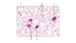
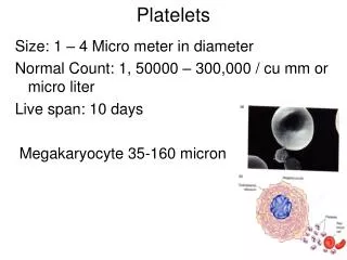
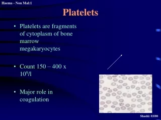
Platelets Platelets are non-nucleated, small, round or oval discs. They are formed in the bone marrow by fragmentation of the cytoplasm of giant cells called “Megakaryocytes”. Platelet count normally = 150,000-400,000/µl. 10
Function of Platelets • Plays a role in Hemostasis = prevention of blood loss. • Whenever a vessel is severed or ruptured, Hemostasis is achieved by several mechanisms; • Vascular spasm. • Formation of a platelet plug. • Formation of a blood clot as a result of blood coagulation.
Platelet Functions Begins with Platelet activation
Platelet Activation • Adhesion • Shape change • Aggregation • Release • Clot Retraction
Platelet function Adhesion Aggregation TxA2 ADP Secretion
Platelet Adhesion • Platelets stick to exposed collagen underlying damaged endothelial cells in vessel wall
Platelet Release Reaction • Platelets activated by adhesion • Extend projections to make contact with each other • Release Thromboxin A2, Serotonin & ADP activating other platelets • Serotonin & Thromboxin A2 are vasoconstrictors decreasing blood flow through the injured vessel. ADP causes stickiness
Platelet Aggregation • Activated platelets stick together and activate new platelets to form a mass called a Platelet Plug • Plug reinforced by fibrin threads formed during clotting process
Platelet • Adhesion Endothelium 2. Shape change • 3. Aggregation
Resting platelet Activated platelet
Platelet Plug Formation Blood vessel injury. Platelets come in contact with damaged blood vessel. They become activated and adhere to the injured area. This will activate even more platelets to come to the injured area resulting in platelet plug formation.
Platelet Plug formation Cont. The platelet plug is a loose plug that is usually successful in blocking the blood loss if the vascular opening is small. Then, during the process of blood coagulation, the stronger fibrin threads are formed that will strengthen the platelet plug.
Platelet Plug Aggregation of platelets at the site of injury to stop bleeding • Exposed collagen attracts platelets • Activated platelets release of platelet ADP & TXA2the stickiness of platelets Platelets aggregation plugging of the cut vessel • Intact endothelium secret prostacyclin inhibit aggregation
Activated Platelets Secrete: • 5HT vasoconstriction • Platelet phospholipid (PF3) clot formation • Thromboxane A2 (TXA2) is a prostaglandin formed from arachidonic acid Function: • vasoconstriction • Platelet aggregation (TXA2 inhibited by aspirin)
Blood coagulation • Initiation on blood coagulation occurs by two ways: • The extrinsic pathway: initiated by trauma to blood vessel. • The intrinsic pathway: initiated in the blood itself.
The Intrinsic Pathway Blood trauma or contact with collagen The Extrinsic Pathway Tissue trauma F XII F XIIa HMW Kininogen Prekallikrein Tissue factor (TF) FXI FXIa Ca++ FVIIa FVII F IX F IXa Ca++ FVIII PLTs Ca++ F X F Xa Blood clot FV PLTs, Ca++ Prothrombin Thrombin FINAL COMMON PATHWAY Fibrin Fibrinogen
Bleeding Disorders • Excessive bleeding can result from; • Platelet defects: deficiency in number (thrombocytopenia) or defect in function. • Deficiency in coagulation factors (e.g. hemophilia). • Vitamin K deficiency.
Bleeding Disorders Cont… • Hemophilia: • ↑ bleeding tendency. • X-linked disease. • Affects males. • 85% due to FVIII deficiency (hemophilia A), and 15% due to FIX deficiency (hemophilia B). • Vitamin K deficiency & liver disease: • Almost all coagulation factors are synthesized in the liver. • Prothrombin, FVII, FIX, & FX require vitamin K for their synthesis.
The Intrinsic Pathway Blood trauma or contact with collagen The Extrinsic Pathway Tissue trauma F XII F XIIa HMW Kininogen Prekallikrein Tissue factor (TF) FXI FXIa Ca++ FVIIa FVII F IX F IXa Ca++ FVIII PLTs Ca++ Blood clot Hemophilia F X F Xa FV PLTs, Ca++ Prothrombin Thrombin Fibrin Fibrinogen
Anticoagulants • Heparin • Liver, lungs, mast cells, basophils • Direct antithrombin • Prevent the conversion of Prothrombin to Thrombin • Injection only • 6-8 hours • Warfarin • Almost all coagulation factors are synthesized in the liver. • Suppresses the synthesis of Prothrombin, FVII, FIX, & FX vitamin K dependent factors • Orally • 48 hours