Download
1 / 28
310 likes | 707 Views
Metabolism of leukocytes and platelets. mirka.rovenska@lfmotol.cuni.cz. Differentiation of the bone marrow stem cells. stem cell. myeloid progenitor. lymphoid progenitor. megakaryocyte. erythroblast. erythrocyte. monocyte. platelets. neutrophil eosinophil basophil. macrophage.

E N D
Metabolism of leukocytes and platelets mirka.rovenska@lfmotol.cuni.cz
Differentiation of the bone marrow stem cells stem cell myeloid progenitor lymphoid progenitor megakaryocyte erythroblast erythrocyte monocyte platelets neutrophil eosinophil basophil macrophage dendritic cell plasma cell mature lymphocytes
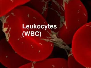
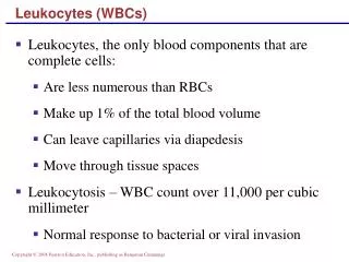
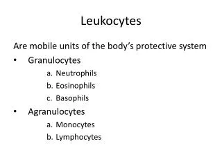
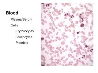
A) Phagocytic cells: • Neutrophils – most abundant • Eosinophils • Monocytes • Macrophages – rise by differentiation of monocytes in tissues
Degradation of the ingested particle: • 1) Activation of NADPH oxidase • 2) Production of NO by nitric oxide synthase • 3) Fusion of phagosome with lysosomes of the phagocytic cell that contain bactericidal substances and hydrolytic enzymes (often with acidic pHopt)
1) NADPH-oxidase • Protein complex of neutrophils, eosinophils, monocytes, macrophages • NADPH + 2 O2→ NADP+ + H+ + 2 O2•- 2 O2•- + 2 H+ → O2 + H2O2 • H2O2 can damage bacteria directly or after conversion to OH• : H2O2 + M+ → OH• + OH- + M2+(M; metal) superoxide anion
Activation: by association of the components localized in cytosol with cytochrome b558 in the membrane; electrons from cytosolic NADPH are – via FAD and cytochrome – transferred to oxygen cytochrome b558 active NADPH-oxidase
plasma membrane fusion with lysosomes phagosome
Myeloperoxidase • Present in granules of neutrophils and monocytes, but not macrophages! • Significant portion of H2O2 (produced by dismutation of O2•- generated by NADPH oxidase) is used by myeloperoxidase to oxidize Cl- to HClO • HClO is highly reactive, able to oxidize biomolecules; it also provides toxic chlorine gas: HClO + H+ + Cl-→ Cl2 + H2O • HClO also reacts with O2•- yielding OH•: HClO + O2•- → O2 + OH• + Cl-
Chronic granulomatous disease • Caused by a deficiency of one of the NADPH oxidase subunits • Superoxide and the other reactive oxygen species are not produced • Severe infections that are very hard to treat – e.g.: • Burkholdaria cepacea causes pneumonia • Aspergillus causes intractable pneumonia, septicaemia; can lead to death • Treatment: antibiotics, antifungal agents
2) Nitric oxide production • Mainly by inducible nitric oxide synthase (iNOS) of macrophages which is induced by cytokines (INF-γ, TNF)or bacterial lipopolysaccharide: • NO• can kill bacteria directly (e.g. by inhibition of the respiratory chain) or indirectly: by reaction with O2•-, generating peroxynitrite ONOO- which attacks Fe-S proteins and essential –SH groups, inactivates enzymes… Arg citrulline
NADPH oxidase is effective mainly in degradation of extracellular pathogens (Salmonella, Staphylococcus, Streptococcus pyogenes)…neutrophils X • NO serves mainly to kill the intracellular parasites (Listeria, Brucella, Candida albicans)…macrophages
3) Granules (lysosomes) of neutrophils • Contain bactericidal substances and hydrolases that, after fusion with phagosome, destroy the engulfed particles: • myeloperoxidase • lysozyme – cleaves glycosidic bonds in peptidoglycan of the bacterial (primarily G+) cell walls • defensins – cationic peptides (Arg) with Mrof 3,5-6 kDa; interact with anionic lipids of bacterial membrane and make pores in it; can also inhibit synthesis of DNA and proteins • hydrolases,e.g.elastase – serine protease: can damage bacteria and cleave virulence factors, but also cause harm to host tissues (cleaves the proteins of extracellular matrix, too)
Eosinophils • Main task: defence against multicellular parasites • Display all the above-mentioned mechanisms with slight differences: • ROS production • peroxidase of eosinophils – similar to myeloperoxidase, but prefers Br-as a substrate (instead of Cl-), thus generating HBrO (instead of HClO) • basic protein of eosinophils disrupting the parasite cell membranes
B) Basophils and mast cells • Activated by antigens / allergens interacting with IgE bound to the surface IgE receptors of basophils (mast cells) • Upon activation, content of their granules is released – substances that are harmful to parasite and induce reactions that should lead to its removal; however, they can also be responsible for allergic symptoms: • hydrolases • histamine • heparin • Synthesis of eicosanoids is activated; leukotrienes are potent bronchoconstrictors, stimulate chemotaxis and leukocyte activation cytoplasmic granules
Histamine • Produced by histidine decarboxylation: • Causes vasodilationand bronchoconstriction helps to eliminate parasites (cough, peristalsis, enhanced production of mucus)
Atopy • IgE recognizing allergens (from pollen, food…) are produced and bind toIgE receptors of basophils (mast cells). Next exposure to the allergen can lead to release of histamine and heparin and synthesis of eicosanoids • Local symptoms occur: allergic rhinitis, asthma, conjunctivitis • If the allergen enters bloodstream, it can cause a massive degranulation of basophils (mast cells) increase in vascular permeability, decrease in blood pressure pulmonary oedema, ischemia… anaphylactic shock • Treatment: antihistamines – block histamine receptors
C) Lymphocytes • Have specific receptors recognizing one particular antigen: B cell receptors (BCR) and T cell receptors (TCR), respectively • BRC is a membrane-bound immunoglobulin, TCR is very similar to Ig • B cells (after proliferation and differentiation into plasma cells) secrete large amounts of antibodies (soluble immunoglobulins)
VH VL CH1 Fab CL CH2 Fc CH3 Soluble immunoglobulins • 2 heavy chains (H) interconnected by disulfide bonds • 2light chains(L), each connected to one of the H chains (by disulfide bond) • H chain: 4-5 domains, 50-75 kDaL chain: 2 domains, 25 kDa • N-terminal domains of H- and L-chains are variable (VH resp. VL), the others are constant (CH resp. CL), i.e. the same in one type of Ig • Variable domains of H a L chains form theantigen-binding site
Types of immunoglobulins (Ig) • There are 2 isotypes of L: κ, λ • There are 5 isotypes of H: , γ, δ, ε, μ • According to these isotypes of H, 5 types of immunoglobulins can be distinguished: • IgA (2 subtypes) • IgG (4 subtypes) • IgD • IgE • IgM • IgM can form pentamer, IgA can form dimer or trimer 155 kDa (similar: IgD, IgE) 900 kDa
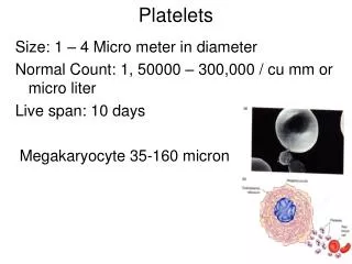
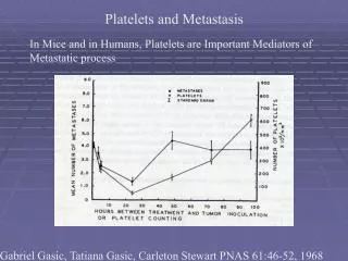
D) Platelets • No nucleus many of their metabolites come from megakaryocytes • Form blood clots, act as vasoconstrictors • Participate in defence against infections, e.g.: they suppress the growth of Plasmodium falciparum (infectious agent that causes malaria) • Generate O2•- and H2O2 that may synergize with pro-aggregatory stimuli • Contain thromboxan A synthase that catalyzes conversion of prosta-glandin H2 to thromboxan A2: TXA2 – promotes platelet aggre- gation and vasoconstriction
Platelets also release two very important factors that can influence not only platelets but also other cell types: • Platelet-Activating Factor (PAF) • Platelet-Derived Growth Factor (PDGF)
Platelet-Activating Factor • Mainly juxtacrine and paracrine signalling via GPCR • Promotes platelet aggregation • Induces activation of leukocytes, adhesion, chemotaxis, cytokine production, causes vasodilation and bronchoconstriction • Mediates interplay between thrombotic and inflammatory cascades • BUT: it is also suspected of contributing to allergy, anaphylactic shock… • It is produced also by endothelial cells, monocytes, granulocytes… phospholipid
Platelet-Derived Growth Factor • Dimeric protein, 3 isoforms • Receptors: tyrosine kinases – expressed on fibroblasts, glia, smooth muscle cells, leukocytes…. • Effects: • proliferation • chemotaxis • cytoskeletal rearrangements • differentiation of certain types of cells (e.g. in CNS) • participates in wound healing, capillary formation, embryonic and postnatal development! • BUT: probably also plays a role in pathogenesis (some tumours)
Cytokines • Proteins secreted by leukocytes and other cells(but there are also membrane cytokines) that influence (via receptors) the cells of the immune system • Cytokine signalling: • autocrine – a cytokine influences the same cell that produces it • paracrine – a cytokine influences the nearby cells • endocrine– a cytokine influences distant cells (after transport by the bloodstream)
Types of cytokines • Interleukins – e.g. IL-6: produced by macrophages, neutrophils, stimu-lates lymphocytes, secretion of Ig, synthesis of acute phase reactants • Chemokines –induce chemotaxis • Interferons – e.g. INF-: produced by lymphocytes, monocytes, and macrophages, participates in antiviral defense (induces synthesis of enzymes that block viral replication) • Transforming growth factors – e.g. TGF-β: produced by T-lymphocytes, macrophages, and platelets, displays anti-inflammatory effects • Tumor necrosis factors – e.g. TNF-β: able to induce apoptosis
Leukocyte infiltration into tissues= diapedesis (extravasation): Taken from: Halliwell, Gutteridge, Oxford University Press, 1999 • Leukocytes are slowed down by the interaction of their mucinswith selectineson the surface of endothelial cells (EC) • Cytokines on the surface of EC interact with the receptors of leukocytes • A strong adhesion mediated by the interaction of integrins with molecules on the surface of EC→ migration of leukocytes into the tissue directed by cytokins released by inflammatory cells or EC
Regulation • Many functions of leukocytes are regulated by monomeric GTP-binding proteins, e.g. Rac, Rho: • activation of NADHP oxidase • chemotaxis • phagocytosis • fusion of phagosome with granules • Rho and Rac are able to modulate the assembly of actin filaments, which plays a role in the processes listed above • http://uk.video.search.yahoo.com/video/play?ei=UTF-8&fr=yfp-t-702&p=chemotaxis&vid=0001539076618&dt=&l=77&turl=http%3A%2F%2Fyts.video.search.yahoo.com%2Fimage%2F2021a80a1&rurl=http%3A%2F%2Fwww.youtube.com%2Fv%2FZUUfdP87Ssg%26hl%3Den%26fs%3D1&tit=Neutrophil