Download
1 / 37
390 likes | 700 Views
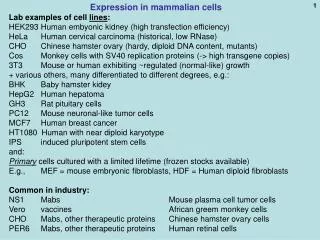
Expression in mammalian cells Lab examples of cell lines : HEK293 Human embyonic kidney (high transfection efficiency) HeLa Human cervical carcinoma (historical, low RNase) CHO Chinese hamster ovary (hardy, diploid DNA content, mutants)

E N D
Expression in mammalian cells Lab examples of cell lines: HEK293 Human embyonic kidney (high transfection efficiency) HeLa Human cervical carcinoma (historical, low RNase) CHO Chinese hamster ovary (hardy, diploid DNA content, mutants) Cos Monkey cells with SV40 replication proteins (-> high transgene copies) 3T3 Mouse or human exhibiting ~regulated (normal-like) growth + various others, many differentiated to different degrees, e.g.: BHK Baby hamster kidey HepG2 Human hepatoma GH3 Rat pituitary cells PC12 Mouse neuronal-like tumor cells MCF7 Human breast cancer HT1080 Human with near diploid karyotype IPS induced pluripotent stem cells and: Primary cells cultured with a limited lifetime (frozen stocks available) E.g., MEF = mouse embryonic fibroblasts, HDF = Human diploid fibroblasts Common in industry: NS1 Mabs Mouse plasma cell tumor cells Vero vaccines African greem monkey cells CHO Mabs, other therapeutic proteins Chinese hamster ovary cells PER6 Mabs, other therapeutic proteins Human retinal cells
Mammalian cell expression Generalized gene structure for mammalian expression: polyA site intron Mam.prom. 3’UTR cDNA gene 5’UTR Intron is optional but a good idea
SV40 LargeT Ag: Simian Virus 40 RSV LTR: Rous sarcoma virus MMTV: Mouse mammary tumor virus, glucocorticoid [Dex] inducible HSV TK: Herpes simplex virus, low expression Metallothionein: many sources, metal inducible, Cd++ CMV early: Cytomegalovirus, strong in most cell types Engineered inducible / repressible:tet-, ecdysone-, glucocorticoid- responsive (tet = tetracycline) Popular mammalian cell promoters
Tet-OFF Engineered regulated expression: Tetracycline-reponsive promoters Tet-OFF (add tet shut off) VP16 tc’nact’n domain (Herpes virus) tetRdomain tTA = tet activator fusion protein: active If no tet,binds tet operator(if tet not also bound) Tet bound, allosteric change in conformation,cannot bind operator, not active Tet-OFF VP16 tc’nact’n domain tetRdomain Tetracycline (tet), or,better, doxicyclin (dox) tTA gene must be in cell (permanent transfection, integrated): polyA site CMV prom. tTA cDNA (Bujold et al.)
Tet operator-repressor, original bacterial source state tet prom. Tetracycline resistance gene RNA pol tet operator sequence No doxicyclin: tetRprotein inactive no transcripton, RNA Pol blocked RNA pol tet prom. Tetracycline resistance gene tet operator sequence tetRprotein Doxicyclin present: active transcripton, no blockage RNA pol tet prom. your favorite gene
tetRdomain VP16 tc’nact’n domain not active little transcripton (2%?, bkgd) Doxicyclin present: polyA site MIN. CMV prom. your favorite gene polyA site polyA site your favorite gene your favorite gene No doxicyclin: VP16 tc’nact’n domain tetRdomain active Plenty of transcripton RNA po l MIN. CMV prom. Tet-OFF, exploits modulatable binding of the tet protein bytet MIN. CMV prom. Mutliple tet operator elements
Tet-ON Tetracycline-reponsive promoters Tet-ON: add tet turn on gene tetRdomain VP16 tc’nact’n domain not active Different fusion protein: Does NOT bind tet operator(if tet not bound) tetRdomain VP16 tc’nact’n domain active Tetracycline (tet), or,better, doxicyclin (dox): Now, can bind to operator seq. polyA site Full CMV prom. tTA cDNA tTA must be in cell (permanent transfection, integrated): commercially available (293, CHO) or do-it-yourself
polyA site polyA site polyA site your favorite gene your favorite gene your favorite gene Tet-ON MIN. CMV prom. Mutliple tet operator elements tetRdomain VP16 tc’nact’n domain not active little transcription (bkgd.) Doxicyclin absent: MIN. CMV prom. Add dox: active tetRdomain VP16 tc’nact’n domain doxicyclin active Plenty of transcripton (> 50X) RNA pol II MIN. CMV prom.
Back to protein-protein interactions: Reporterenzyme Enzyme fragments themselves do not associate well enough to reconstitute an active enzyme F = reporter protein fragment SW Michnick web site: http://michnick.bcm.umontreal.ca/research/images/pca_general_en.gif
Dihydrofolate reductase (DHFR): role in metabolism Folic acid DHFR (FH2) DHFR (FH4) http://www.nature.com/onc/journal/v22/n47/images/1206946f1.gif
Clonal selection and in vivo quantitation of protein interactions with protein-fragment complementation assays, I. Remy and S.W. Michnick PNAS 96, 394–5399, 1999 DHFR fragments Rapamycin promotes the association of the 2 protein domains fMTX Cell growth assay: CHO DHFR- mutantcells Fluorescein – MTXbinding assay IN PURINE-FREE MEDIUM DHFR = dihydrofolate reductase DHF=dihydrofolate = FH2 THF=tetrahydrofolate = FH4 fMTX=fluorescent methotrexate FK506 = immunosuppressant drug FKBP = FK506 binding protein FRAP = FKBP–rapamycin binding proteinFRB= FKBP–rapamycin binding domain of FRAP
FK506 recruits FKBP to bind to calcineurin and inhibit its action as a specific phosphatase a phosphatase
Claim detection of 0.05 nM rapamycin ?? No. of CHO colonies [rapamycin]
Fluorescent methotrexate (fMTX) assay: Wash in, wash out CHO cells (permanent transfection) cos cells (transient transfection) Leucine zipper protein fragments instead of rapamycin binding proteins (positive contro) Background association of FKBP and FRB without rapamycin (compare mixed input)
Fuorescence-activated flow cytometer (FACS is this, plus more) Allows quantitation of fluorescence per cell No. of cells 8-fold increase in fluorescence per cell Log of fluorescence intensity Fluorescence intensity Measure affinity for a drug in vivo [rapamycin] Competition with a molecule that binds only one
Erythropoietin-erythropoietin receptor (dimer) interaction: Efficacy of a peptide mimetic EPO EPO bp2 EPO bp1 Erytropoietin (EPO) receptor In vivo assay of drug effectiveness (EMP1) (inexpensive substitute for erythropoietin?) EMP1 = Erythropoietin mimetic peptide 1 Erythropoietin
FACS = Fluorescence-activated cell sorter Impart a charge on the recognized cell Less than one cell or particle per droplet. Thus the most that most droplets contain is one particle. Can be used purely anaytically without the sorting capability. Then called “flow cytometry”, or also called FACS anyway. Charged plates attract droplets containing a particle of the opposite charge Cells remain viable if treated with care.
Histogram-type display No fluorescence (background autofluorescence) No. of cells Red stained Usually a log scale Having this much fluorescence
Scatter plot display Analysis on 2 colors One cell Amount of greenfluorescence (log) You decide on the positions of of demarcations Say, want high reds but low greens: Instruct the FACS to deflect cells in this quadrant only. Collect and grow or analyze further. Amount of red fluorescence (log)
Beaming bead FACS analysis Analysis of beads representing the human genome using allele-specific hybridization probes and the FACS A. Flow cytometry data: 2-D plots where each point represents one particle. Then contour lines plotted around the point density. Here light “forward” scattering (irrespective of wavelength) is measured (FSC). Instrument can be set to reject data from 2-bead doublets that scatter light more. Both signals Red signal Neither signal Green signal B-D. Amplified beads hybridized to 2 probes, one specific to the S allele of a certain gene and one specific to the L allele. The beads carry the amplified PCR products corresponding to this region from 3 human individuals. The blue points come from microspheres that contained both types of PCR products from both alleles, despite the high dilution.
Biotechnology methods to study transcriptional regulation in cells Mainly, use of reporter proteins whose cDNA sequence is linked to the promoter. First, a synopsis of promoter structure:
General model for transcriptional regulation in higher eukaryotes Core transcriptional elements TF… transcription factor TBP: TATA binding protein TAF: TBP associated protein BRE: TFIIB response element INR: transcription initiator element DPE: downstream promoter element -28 -35 GGGCGCC; CCACGCC YYAN(TA)YY (AG)G(AT)(CT)(GAC) TATA(AT)AA(GA) Y = C or T (pyrimidine) The transcription complex either recruits RNA Pol II or activates a bound RNA Pol II For review see Smale and Katonga, Ann. Rev. Biochem. 72: 449-479 (2003)
Many transcriptional enhancer elements often lie upstream of promoters,allowing for many combinations of TF binding
Put a DNA regulatory region upstream of a reporter gene to analyze its elements Space for res. enz. to bind Reportergene PCR Transfect
Beta-galactosidase (β-gal) – detection by several different assays Chloramphenicol acetyl transferase (CAT) – detection, sensitive radioactive assay Luciferase (firefly, Renilla [jellyfish]) – detection, easy dual, sensitive luminescent assay Green fluorescent protein (GFP, BFP, YFP)) – cytological, visible in living cells, fusion proteins, FACS Neomycin phosphotransferase (neo)–selectable drug resistance (G418R) (similarly: resistance to hygromycin, puromycin, histidinol Dihydrofolate reductase (DHFR) – selectable in dhfr- cells, amplifiable, fusion proteins work Suicide selection: Herpes simplex virus thymidine kinase (HSVTK) Popular reporters to study promoter/enhancers FACS = fluorescence-activated cell sorter
Gangcyclovir selection AGAINST the presence of enzyme activity (compare to 5-fluoro-orotic acid (FOA) resistance in yeast, URA3-) CRE recombinase (cassette excnahge) Mut. protein of interest HSVTK Gancilovir, ATP Gancilovir-PO4 toxicity, death Mammalian TK Gangcylovir, ATP (Ganciclovir itself is not toxic) Use example: Site-directed recombination Engineered chromosome: lox lox WT protein of interest HSVTK Replacement plasmid: gangcylovir Mut. protein of interest Select recombinants as HSVTK-, gancilovir-resistant
Testing for a cell-specific promoter: chloramphenicol acetyl transferase (CAT) reporter assay CAT cDNA is from a prokaryotic source. CAT is not found in mammalian cells. Therefore low backgrounds diacetylated B A Thin layer chromatography (TLC) 14C-chloramphenicol monoacetylated unacetylated Positive control Negative control
ONPG (ortho-nitrophenyl-beta-galactoside) – spectrophotometric measurement (420 nm – blue color – simplest) X-gal (5-Bromo-4-chloro-3-indolyl-ß-D-galactoside) – blue precipitate - for cytology or colony detection Umbelliferyl–galactoside (-> umbelliferone, fluorescent, reading in a fluorimeter allows more sensitive quantification than spectrophotometry) Galacton-STAR or some such (-> chemiluminescent product = emission of light, so lower background than fluorescence) Lactose (glucose-beta-galactose disaccharide) – allows growth if hydrolyzed; growth phenotype. For microbial cells usually. Reporter enzyme substrates for different purposes Substrates for beta-galactosidase, for example:
Light units of luciferase in hepatocytes Mapping transcriptional elements upstream of a promoter: Mapping with restrictionenzyme mediated deletions Conclusion:
Footprinting: detects sites on DNA to which protein are bound DNA + DNA-binding protein Naked DNA Population of molecules Partial DNase Population of molecules missing Gel electrophoresis.autoradiography Footprint
Protein-DNA binding: EMSA or gel shift (EMSA = electrophoretic mobility shift assay) 1 2 3 4 5 competitor (supershift) (shift) DNA element (Even though the hexagon looks like a protein here) U. Arizona
Gel shifts (EMSA (surpershifted complex is not competed by NON-specific probe) Protein DNA complexes migrate more slowly than naked DNA (competed only by specific probe) Super- shift (two molecules of protein bound)
SELEX for protein binding sites Systematic Evolution of Ligands by Exponential Enrichment (T7 RNA Pol from an embedded T7Pol promoter (huge number) Synthetic, range usually 6 to 40-mers (usually a protein) ; by PCR (re-iterate 3-10 times) Binding to Protein, e.g. Separate using nitrocellulose binding, gel electrophoresis, etc. sequences consensus
Practical capacity ($700): 1014 random sequences (random ~21-mer = 421) Binding to protein of interest http://www.molmed.uni-luebeck.de/T.%20Restle/Bilder/SELEX.jpg RT
Binding site for a “puf “ protein, implicated in mRNA degradation PUM2, a novel murine puf protein, and its consensus RNA-binding siteWhite EK, Moore-Jarrett T, Ruley HE. RNA. 2001 Dec;7(12):1855-66. 20-mer Nucleic acid degenerate base abbreviations Consensus: Description