Download
1 / 9
90 likes | 383 Views
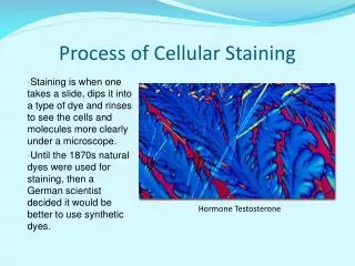
Observation of bacteria using staining procedures. Simple staining Gram staining. Simple Staining. Smear preparation: A drop of water is placed in the centre of a slide One loopfuls of organisms is transferred to the centre of slide Spread the organisms over the slide

E N D
Observation of bacteria using staining procedures Simple staining Gram staining
Simple Staining • Smear preparation: • A drop of water is placed in the centre of a slide • One loopfuls of organisms is transferred to the centre of slide • Spread the organisms over the slide • The smear is allowed to dry • Slide is passed through flame several times to heat-kill and fix organisms • A bacterial stain is stained with crystal violet (fuchsin, methylene blue) 1 min • Stain is briefly washed off slide with water Allow the slide to air-dry and examine with an oil immersion objective
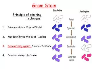
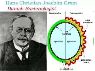
Gram-positive cell wall Gram Staining 1884 Christian Gram Staining technique that separates bacteria into two groups: Gram-positive bacteria Gram-negative bacteria Based on the ability to retain crystal violet during decolorization with alcohol Gram-negative cell wall
1.Step. Fixation, staining with crystal violet 2. Step. Gram`s iodine 3. Step. Ethyl alcohol 4. Step. Counterstaining with fuchsin G+ violet (blue) G- red (pink)
Grampositive bacteria • Steptococcus • Staphylococcus • Lactobacillus • Bacillus • Clostridium
Gram-negative bacteria • Escherichia • Salmonella • Vibrio • Treponema
Gram Staining • Smear preparation. • Stain with crystal violet 1 min. • Add Lugol solution 1 min. • Decolorize with alcohol 10-15 seconds. • Wash with water. • Stain with fuchsin 2 min • Allow the slide to air-dry • Examine with an oil immersion objective