Staining
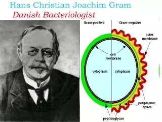
A Danish Bacteriologist developed the staining method

Staining
E N D
Presentation Transcript
Dyes become Stains: • In 1884, Hans Christian Gram, a Danish doctor working in Berlin, a method which still forms the basis for the identification of bacteria. • While examining lung tissue from patients who had died of pneumonia, he discovered that certain stains were particularly taken up and retained by bacterial cells. • Over the course of the next few years, • Gram developed a staining procedure which divided almost all bacteria into two large groups - the Gram stain.
History: • With interest in the effects of dyes on living tissue. In Danishmicrobiologist Hans Christian Gram discovered that crystal violet irreversibly stains certain bacteria but can be washed from others. • The dye has been widely used ever since for the Gram stain technique, which identifies bacteria as gram-positive (the stain is retained) or gram-negative (the stain is washed).
History: • It was later modified by Hucker in 1921. • The modified procedure provided greater reagent stability and better. • Differentiation of organisms. Other modifications have been intentionally. • Developed for staining anaerobes and for weakly staining gram-negative bacilli. • (Legionella spp., Campylobacter spp., Bacteroides spp. Fusobacterium spp.)
Gram staining a Important Technique • A staining technique used to classify bacteria; • Bacteria are stained with violet and then treated with Gram's solution. • After being decolorized with alcohol and treated with safranine and washed in water, those that retain the violet are Gram-positiveand those that do not retain it are Gram-negative.
Gram positive Bacteria: • Gram positive bacteria have a thick cell wall of peptidoglycan and other polymers. • Peptidoglycan consists of interleaving filaments made up of alternating acetylmuramic acid and actylglucosamine monomers. • In Gram positive bacteria, there are "wall teichoic acids".
Gram Negative Bacteria: • Gram negative bacteria have an outer membrane of phospm, holipids and bacterial Lipopolysaccharides outside of their thin peptidoglycan layer. • The space between the outer membrane and the peptidoglycan layer is called the periplasmic space. • The outer membrane protects Gram negative bacteria against penicillin and lysozymes.
Comparison of Gram-Positive and Gram-Negative Cell Walls Characteristic Gram-Positive Gram-Negative Number of major layers 1 2 Chemical composition Peptidoglycan Lipopolysaccharide Teichoic acid Lipoprotein Lipoteichoic acid Peptidoglycan Overall thickness Thicker Thinner (20–80 nm) (8–11 nm) Outer membrane No Yes Periplasmic space Narrow Extensive Porin proteins No Yes Permeability to molecules More penetrable Less penetrable
A method of staining bacteria using a violet stain. • The gram staining characteristics (denoted as positive or negative). • A heat fixed bacterial smear is stained with crystal violet, treated with 3% iodine / potassium iodide solution, washed with alcohol and counterstained. • The method differentiates bacteria into two main classes,gram-positive and gram-negative.
Prefer to pick up colonies with loop and smear on Clean glass slide
Making a Smear: • First prepare your slide. You do this by placing bacteria on a slide in a drop of water, allowing them to dry and then heat fixing them. • Heating the slide kills the bacteria and makes sure that the bacteria a fixed to the slide and wash away during the staining procedure.
Choosing a Right Smear: • Before choosing a field for microscopic examination, it is important to look at the smear macroscopically . • Note that the smear is easily visible in ordinary light .
Requirements for Gram staining technique: a. Glass slides (25 X 75 mm), frosted ends desirable b. 0.85% Nacl, sterile c. Pasteur pipettes and wood applicator sticks, sterile d. Microbiological loops, inoculating needles e. Supplies for disposal of biological waste, including “sharped” f. Bunsen burner g. Immersion oil
Making Multiple smears in same slide conserve resources: • Making multiple smears make the optimal use of the slide. • Reduces the economic costs and saves the technical time.
Correct preparation: • Smear preparation: Proper smear preparation should produce a monolayer of organisms sufficiently dense for easy visualization but thin enough to reveal characteristicmorphological characteristics. Use clean, new glass slides. NOTE: When using the same pipette or swab, always inoculate culture media first
Steps in Gram Staining Procedure: 1.On a rack, overflow with filtered crystal violet 10 sec. 2. Wash briefly in water to remove excess crystal violet. 3. Flood with Gram’s iodine 10 sec. 4. Wash briefly in water, do not allow the section dry out. 5. Decolourise with alcohol until the moving dye front has passed the lower edge of the section 6. Wash immediately in tap water. 7. Counterstain with safranin 15 seconds.
Microscopy: The Instruments • In a compound microscope the image from the objective lens is magnified again by the ocular lens. • Total magnification =objective lens ocular lens
Gram Stain Differentiates Gram positive from Gram negative • Differential Stains: Gram Stain
SPORE STAINING: Detects endospores generated by Bacillus and Clostridium: Formed within the cell dormant strong in adverse conditions Various sizes, shapes and locations Steps(Schaffer-Fulton procedure): • Heat + malachite green • Rinse • Counter stain with saffranin
CAPSULE STAIN: Gently – no heat fixation or may get shrinkage Steps: • Grow culture in skim milk broth • Crystal violet • Copper sulfate counter stain
FLAGELLA STAIN: Flagella are very difficult to observe directly because of their size Thickness is increased using mordant's • Tannic acid • Potassium alum Stained with Pararosaniline (Leifson method) or basic fuchsin (Gray method).
Trouble shooting in Gram Staining method: 1. The age of the culture. Cultures more than 24 hours old may lose their ability to retain the crystal violet-iodine complex. 2. The organism itself. Some gram-positive bacteria are more able to retain the crystal violet-iodine complex than others. One must use very precise techniques in gram staining and understand the results with carefulness.
Modification in Gram staining methods: • Some of them have improved the method, • Bartholomew (1962) has pointed out that each variation in the Gram staining procedure has a specific limit to its acceptability. • Any final result is the outcome of the interaction of all of the possible variables. • All modified methods to be practised with caution to the laboratory and quality control checks.