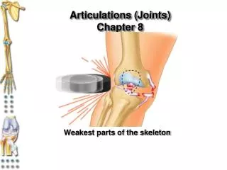
Chapter 8 Joints
Chapter 8 Joints. Structural Classification of Joints Functional Classification of Joints Movements Special Joints Diseases and Disorders. Structural Joint Classification. Fibrous Sutures – better described as synostoses (skull) Syndesmoses – tibia-fibula slight vs. a lot radius-ulna

Chapter 8 Joints
E N D
Presentation Transcript
Chapter 8 Joints • Structural Classification of Joints • Functional Classification of Joints • Movements • Special Joints • Diseases and Disorders
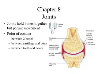
Structural Joint Classification • Fibrous • Sutures – better described as synostoses (skull) • Syndesmoses – tibia-fibula slight vs. a lot radius-ulna • Gomphoses – tooth attachments periodontal ligament • Cartilaginous • Synchondroses – epiphyseal plates and rib-sternum • Symphyses – intervertebral bones and pubic bones where there is fibrocartilage separating the bone. • Synovial
Functional Classification of Joints • Synarthroses – no movement • Amphiarthroses – limited movement • Diarthroses – extensive motion capabilities in mobile joints of limbs
Synovial Joint Characteristics • Articular Cartilage • Joint Synovial) Cavity • Articular Capsule (dense irregular fibrous) with inner layer that is the synovial membrane • Synovial Fluid viscous due to hyaluronic acid • “Weeping Lubrication” thins w/ warming • Reinforcing Ligaments • Capsular or intrinsic ligaments w/in the capsule • Extracapsular outside • Intracapsular sort of “inside”
Bursae & Tendon Sheaths • Protection where tendons “rub” bones • Protection of the tendons
Joint Stability • Articular Surfaces – often only a minor role! • depth • fit • Ligaments • stretch limitations (6%) • Muscle Tone – often THE MOST important • shoulder & knee especially
Movement Direction • Driven by muscles that attach to bones or connective tissue (CT) at, at least, two points • Muscle origin – immovable or less movable bone • Insertion – more movable bone *contraction moves the insertion closer to the origin • Nonaxial • Uniaxial • Biaxial • Multiaxial
Type of Movements of Synovial Joints • Gliding a.k.a. translational • carpus and tarsus, and articular processes of vertebrae • Angular ↑ or ↓ angle between bones • Flexion/Extension • Dorsiflexion/Plantar Flexion • Abduction/Adduction • Circumduction • Rotation – C1 to C2 or the radius or lateral or medial movement of the hip • Special Movements
Types of Synovial Joints • Plane • Hinge • Pivot • Condyloid • Saddle • Ball-and-Socket
Condyloid or Ellipsoidal Joints • Oval articular surface of one bone fits into a complementary depression in another • Both articular surfaces are oval • Biaxial joints permit all angular motions • Examples: radiocarpal (wrist) joints, and metacarpophalangeal (knuckle) joints
Condyloid or Ellipsoidal Joints • Oval articular surface of one bone fits into a complementary depression in another • Both articular surfaces are oval • Biaxial joints permit all angular motions • Examples: radiocarpal (wrist) joints, and metacarpophalangeal (knuckle) joints
Saddle Joints • Similar to condyloid joints but allow greater movement • Each articular surface has both a concave and a convex surface • Example: carpometacarpal joint of the thumb
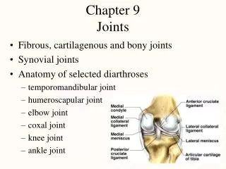
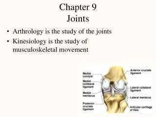
The Knee • Multiple sets of ligaments • Motion allowed in flexion that is prohibited by ligaments when in extension • Tibial Collateral (MCL) • Fibular Collateral (LCL) • Popliteal • ACL/PCL • Capsular ligament support • Patellar Ligament
Synovial Joints: Knee • Largest and most complex joint of the body • Allows flexion, extension, and some rotation • Three joints in one surrounded by a single joint cavity • Femoropatellar joint • Lateral and medial tibiofemoral joints
Synovial Joints: Knee Ligaments and Tendons – Anterior View • Tendon of the quadriceps femoris muscle • Lateral and medial patellar retinacula • Fibular and tibial collateral ligaments • Patellar ligament Figure 8.8c
Synovial Joints: Knee – Posterior Superficial View • Adductor magnus tendon • Articular capsule • Oblique popliteal ligament • Arcuate popliteal ligament • Semimembranosus tendon Figure 8.8e
The Shoulder • Tremendous flexibility & range of motion • Limited structural support • Shallow glenoid fossa • Minor capsular ligaments • Labrum • “Rotator Cuff”
Rotator Cuff Muscles • Long head of Biceps - stabilizes • Supra & Infraspinatus • Teres Minor • Subscapularis
The Hip (Os Coxae) • Highly mobile • More ligamentous support than shoulder • Deeper articular fossa in acetabulum • Labrum diameter < femoral head • Heavy musculature and tendons surround
The Elbow • Radial Collateral Ligament (Lateral) • Ulnar Collateral Ligament (Medial) 3 parts a.k.a. “Tommy John” surgery • Radial Annular Ligament • Articular Capsular Ligaments
Common Joint & Movement Problems • Sprains & strains – stretch or tear support ligaments w/ ankle, knee, shoulder and vertebrae common sites. Poor healing due to poor blood supply, and have lots of pain and immobility. Need surgery so ligament lasts • Cartilage injuries – most often is meniscal tears, but a big increase in young athletes joint surface tears. These are avascular = no healing. Arthroscopic surgery used • Dislocation vs. “separation” – (luxation vs. subluxation) where bones are out of alignment at a joint, accompany sprains. Jaw fingers/thumb, and shoulder – must reduce • Bursitis – inflammation in a bursa, often with or w/o tendonitis. Trauma and friction (abuse) causes fluid accumulation. Tx: cortisone, rest, ice, NSAID’s
Cont. • Arthritis – joint inflammation, 100 different types of degenerative joint disease and inflammation! Crippling painful and stiff, w/ swelling when acute. Infections, trauma, wear and tear, etc. • Osteoarthritis – age-related wear, but they may be genetically predisposed. NSAID’s → Surgery. Glucosamine and chondroitin sulfate may help preserve cartilage, & ↓ crepitus • Rhuematoid – chronic inflammatory disease, ♀ more than ♂, 40-50 yr. olds, some much earlier. Synovitis → pannus formation on the joint margins. Bilateral and in many joints: fingers, wrists, ankles, & feet. Often occurs with exacerbation (relapse) and remission. Believed to be an autoimmune disease that may be related to bacteria. Enbrel, Remicade, Viox, Celebrex have been used to treat (Viox off the market) • Gout – uric acid in blood causes urate crystals in joints, more in ♂ (estrogen↑ excretion), related to diet, often in big toe