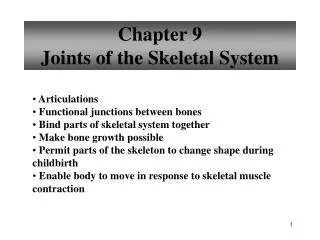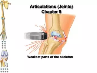
Chapter 9 Joints
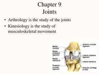
Chapter 9 Joints. Lecture slides prepared by Curtis DeFriez , Weber State University. Joint Classification. Joints have both structural and functional classifications:


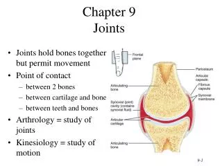
Chapter 9 Joints
E N D
Presentation Transcript
Chapter 9Joints Lecture slides prepared by Curtis DeFriez, Weber State University
Joint Classification • Joints have both structural and functional classifications: • The criteria for classifying joints structurally are anatomical: The presence or absence of a space between the articulating bones and the type of C.T. that binds the bones together. • Functional classification relates to the degree of movement they permit.
Joint Classification • Structural classification subcategories include: • Fibrous joints (bones held together by dense collagen fibers) • Cartilaginous joints (bones held together by cartilage) • Synovial joints (bones held together by ligaments) • Functional classification subcategories include: • Synarthrosis (an immovable joint) • Amphiarthrosis (a slightly movable joint) • Diarthrosis (a freely movable joint)
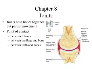
Classification by Structure • Fibrous joints lack cartilage and a synovial cavity. • The bones are held closely together by dense irregular connective tissue. • Suture joints in the skull and the teeth joints
Classification by Structure • Cartilaginous jointsconsist of a bar of cartilage between two bones. • They lack a synovial cavity and provide little or no movement. • Pubic symphysis and the intervertebral disks of the spine
Classification by Structure • Synovial joints are more complex than the other two: Ligaments hold bones together to form a synovial cavity and a freely moveable joint. • A two layered capsule encloses the synovial cavity: • An outer fibrous capsule • An inner synovial membrane
Classification by Structure • The synovial membrane secretes synovial fluid which functions to reduce friction by lubricating the joint and absorbing shocks. It also supplies oxygen and nutrients to the cartilage, while removing carbon dioxide and metabolic wastes. • The major joints of the arms, hips, and legs
Classification by Function • Synarthroses are immoveable joints, like the fibrous joints of the skull. • Amphiarthroses are slightly movable joints like the cartilaginous pubic symphysis. • Diarthroses are freely moveable joints like the big “ball and socket” synovial joints of the shoulder and hip.
Synovial Joints • Because the synovial joint is the most complex, we will look at it now in more detail, including the accessory structures. • Synovial joints are surrounded by accessory structures like the joint capsule, ligaments, and sometimes bursae.
Synovial Joints Accessory structures • Joint capsules are composed of dense irregular C.T., lined by a synovial membrane. • They encompass the joint cavity and the synovial fluid within it.
Synovial Joints Accessory structures • Ligaments are bands of dense regular C.T. (like tendons) that join one bone to another bone. • Bursae (and tendon sheaths) are fluid-filled structures strategically placed to minimize friction in some joints.
Synovial Joints • Accessory structures • In some joints where hyaline cartilage predominates, pads of dense fibrocartilage called menisci are also found between the articular surfaces. • These “articular discs” provide superior strength and allow bones of different shapes to fit together more tightly.
Synovial Joints Accessory structures • Notice that ligaments can blend with other C.T. to become part of a joint capsule, or they can run inside or outside the joint. • The ACL (ligament) lies inside the knee joint, whereas the patellar ligament is outside the joint.
Synovial Joints Accessory structures • Nerve and Blood Supply • Arterial branches from several different arteries merge around a joint before penetrating the articular capsule. • Nerve endings respond to the degree of movement and stretch, and convey information about pain from the joint to the spinal cord and brain.
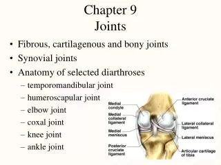
Synovial Joints Accessory structures of the right shoulder joint, anterior view
Types of Synovial Joints • There are 6 types of synovial joints based on the shapes of the articulating bone surfaces. • Not all synovial joints have all (or any) accessory structures like ligaments and bursae – some of them are quite simple.
Types of Synovial Joints • In a planar joint,the articulating surface is flat or slightly curved, permitting back and forth and side-to-side movements.
Types of Synovial Joints • In a hinge joint,the convex surface of one bone fits into the concave surface of another, producing an opening and closing action like a hinge.
Types of Synovial Joints • In a pivot joint, the rounded surface of one bone articulates with a ring structure formed by another bone and a ligament (allowing rotation around its longitudinal axis).
Types of Synovial Joints • In a condyloid joint,the convex oval-shaped projection of one bone fits into the oval-shaped depression of another bone (allowing movement around two axes).
Types of Synovial Joints • In a saddle joint,the articular surface of one bone is saddle-shaped. This is really a modified condyloid joint, but the range of motion is expanded to include movement around all 3 axes.
Types of Synovial Joints • In a ball-and-socket joint,the ball surface of one bone fits into a cuplike depression of another bone. These joints allows the most movement of any joint. • The shoulder joint is a ball-and-socket synovial joint – it has the most range of motion of any joint in the body.
Joint Movements • Range of motion (ROM) refers to the range, measured in degrees of a circle, through which the bones of a joint can be moved. Some of the factors that contribute to keeping the articular surfaces in contact (and affect ROM) include: • Structure or shape of the articulating bones • The shape of the articulating bones determines how closely they fit together. • The strength and tension of the muscles and joint ligaments varies to restrict or permit certain positions.
Joint Movements • ROM is also affected by: • Hormones • Relaxin increases the flexibility of the pubic symphysis and loosens the ligaments between the sacrum and hip bone toward the end of pregnancy. • Disuse • Movement may be restricted if a joint has not been used for an extended period.