Chapter 9-- Joints
Chapter 9-- Joints. Famous Quotes. I figured my body always would be able to repair itself. I think all of us believe that – until you begin to age and get hit with deteriorating joints . Lee Majors

Chapter 9-- Joints
E N D
Presentation Transcript
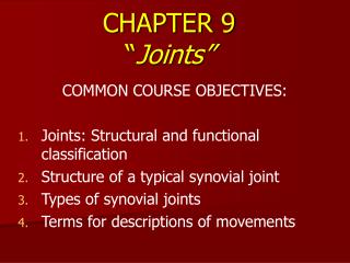
Famous Quotes I figured my body always would be able to repair itself. I think all of us believe that – until you begin to age and get hit with deteriorating joints. Lee Majors It had not occurred to me that marriage requires the same effort as a career. And unlike a career, marriage requires a joint effort. Jessica Savitch (1947-1983)
Ch. 9 (Joints) Study Guide Critically read Chapter 9 pp. 278-297 before 9.3 (Anatomy of selected diarthroses). Study all figures in the PowerPoint slides as well. Comprehend Terminology (those in bold in the textbook) within the reading scope above Study-- Figure questions, Think About It questions, and Before You Go On (section-ending) questions (within the reading scope above) Do end-of-chapter questions— Testing Your Recall— 1-4, 6-7, 9, 11-16, 18 True or False– 2, 4, 5, 9, 10 Review/study review sheet document (4 pages) posted on Blackboard
§ Why Knuckles Crack and Joints Creak Some peoples' bodies play a percussive symphony of cracking and creaking, thanks to the large orchestra of noise-making human joints. What's behind it all?
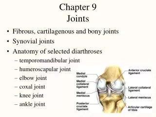
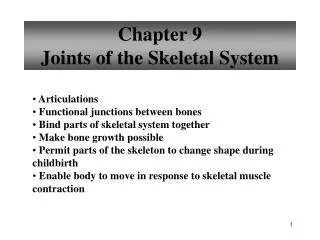
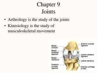
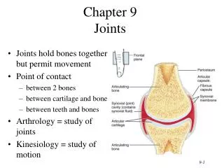
§ Joints and their names • What are joints? • Arthrology = study of the joints • Kinesiology = study of musculoskeletal movement (motion of human body) • How joints are named? • From the names of the bones involved • Ex. The atlantoccipital joint • Ex. The humeroscapular joint
Are all joints able to move? • Immovable joints– where? Why? • Less movable– ex. vertebral column; why? • Moveable—shoulder, elbow, knee • Functional classification (freedom of movement) • synarthrosis (little/no movement) (“Syn”--together) • amphiarthrosis (slightly movable) • diarthrosis (freely movable) (‘Dia”– through)
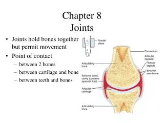
§ Structural classification How adjacent bones are joined? • Bony joints (synostosis; synarthrosis)–two bones fused • Fibrous joints (synarthrosis)—held together by collagen fibers; lack a synovial cavity • Cartilaginous joints (amphiarthrosis)– held together by cartilage; lack a synovial cavity • Synovial joints (diarthrosis)– having synovial (joint/articular) cavity (Fig. 9.5 next slide)
Fig. 9.5 Synovial Joint • Joint in which two bones are separated by a space called a synovial (joint) cavity • Most are freely movable
§ 1. Bony Joints (Synostosis) • Gap between “two bones ossify”– become a single bone • TWO frontal/mandibular bones in infants • Can occur in either fibrous joints or cartilaginous as well; Examples: • Aging: cranial sutures (fibrous joints) destroyed (ex. Parietal bones fuse) in elderly • Aging: attachment of first rib to the sternum (cartilaginous joint) becomes a synostosis with age
§ 2. Fibrous Joints (Synarthrosis-Amphiarthrosis) • Collagen fibers span the space between bones • Three kinds of fibrous joints • A. Sutures— the fibers are short and allow for little movement • B. Gomphoses— the fibers are also _____ and allow for little movement; between teeth and the jaw • C. Syndesmoses—longer fibers and more movable • Figure 9.2
Fibrous Connective Tissue Fig. 9.2 R. Tibia B A Suture Syndesmosis Gomphosis
Fibrous Joint – A. Sutures • Immovable fibrous joints • Bind bones together; only in skull • 3 types— (Fig. 9.3 next slide) • Serrate sutures -interlocking lines • coronal, sagittal and lambdoid sutures • Lap sutures - overlapping beveled edges • Between temporal and parietal bones; Name the suture? • Plane sutures - straight, nonoverlapping edges • palatine processes of the maxillae Suture
Three types of suture-- Lap suture Serrate suture Plane suture Bone Wood Dovetail joint Miter joint Butt joint
Fibrous Joint – B. Gomphoses • Attachment of a tooth to its socket • Held in place by fibrous periodontal ligament • Consisting of collagen fibers attach teeth to jawbones (which bones?) • Little movement (Synarthrosis) while chewing Gomphoses
Fibrous Joint – C. Syndesmosis Tibia & fibula • Two bones bound by broad fibrous sheet called • Interosseous membrane • More/less movable than that of sutures/gomphoses • Examples: radius to ulna (a more movable one, Amphiarthrosis) allow supination and pronation (next slide); tibia to fibula (less movable one) Syndesmosis
Supination and Pronation • For example: In the forearm • Supination • rotation of forearm so that the palm faces forward • Pronation • rotation of forearm so the palm faces to the rear A B Supine means up. In order to carry a bowl of soup, your hand must be in the supine position
Check point question #1-- Functionally, why are sutures classified as synarthroses, and syndesmoses as amphiarthroses? 9-21
§ 3. Cartilaginous Joints (amphiarthrosis) Two bones are linked by cartilage Two types– A. synchondroses and B. symphyses
Cartilaginous Joint – A. Synchondrosis A • Bones are joined by hyaline cartilage • Examples: • First rib attachment to sternum (A on the right) • Temporary joint between epiphysis and diaphysis in growing bones: called Epiphyseal ?__________ B, next
Cartilaginous Joint – B. Symphyses • 2 bones joined by fibrocartilage • Examples: • See figure at right • Only slight amount of movement is possible • Examples– 1 & 2 on the right 2 1
Check point question #2-- What is the structural difference in histology between a synchondrosis and a symphysis? 9-25
§ 4. Synovial Joint • Most are freely movable; two bones in fact Do / Do not touch each other • Two bones are separated by a space called A._________ cavity (with fluid in it) and a soft tissue called B._____________ • B. Articular cartilage -- hyaline cartilage covering the joint surfaces • Synovial fluid —a lubricant; feeds cartilages B A Next slide
§ Synovial joints • Joint (Articular) capsule encloses joint cavity • (Outer) Fibrous capsule: continuous with periosteum • (Inner) synovial membrane; secretes ____________ • Articular discs or menisci: cartilage grows inward and forms pads (Fig. 9.29 c-d; 8.39) • jaw, wrist, sternoclavicular and knee joints • absorbs shock, guides bone movements and distributes forces • Tendon: attaches muscle to bone • Ligament: attaches bone to bone
Right Knee Joint – Superior View Medial meniscus & lateral meniscus absorb shock and shape joint PCL ? ACL Medial condyle of tibia Lateral condyle of tibia
Posterior view Anterior view 9-31
§ Tendon Sheaths and Bursae Ulnar bursa Tendons (flexor digitorum) • Bursa = a sac filled with synovial fluid • Tendon sheaths = elongated bursae lined with synovial membrane and wrapped around a tendon Tendon sheaths
Tendon Sheaths and Bursae Bursa = a sac filled with synovial fluid (Locations) between muscles or between a tendon & a bone etc.; Good examples– Shoulder joint etc. Fig. 9.24 Tendon sheaths = elongated bursae (Locations) where there is considerable friction; such as the hand, wrist, fingers, the ankle, foot, toes etc. Functions of bursa and tendon sheaths: Reduce friction in joints (such as the shoulder), Cushion the movement of one body part over another
Check point question #3-- A) What is the functional classification of synovial joints? B) Why is a meniscus in an interphalangeal joint unnecessary? 9-35
§ 6 Types of Synovial Joints—in descending order of mobility 1.
1. Ball-and-Socket Joints • Features: Smooth hemispherical head fits within a cuplike depression • Examples: • (shoulder) head of humerus into glenoid cavity of scapula • (hip) head of femur into acetabulum of hip bone • Type: only multiaxial joints in the body • Demonstration: (next slide)
Planes of movement (A-C) & axes of rotation A The arm rises in the frontal plane C • Shoulder joint has 3 degrees of freedom = multiaxial joint; • Other joints – monoaxial or biaxial • Axis of rotation– Def.--perpendicular to the plane of movement; examples— A, B, C B The arm rotates in the transverse plane The arm moves through the sagittal plane
Planes of movement & axes of rotation A The arm rises in the frontal plane C • A—Abduction (away from midline) vs. adduction • B—Flexion (decreases a joint angle) vs. extension • C—Medial (internal) rotation vs. lateral rotation B The arm rotates in the ______ plane It moves through the _________ plane
2. Condyloid (ellipsoid) Joints • Features: Oval convex surface on one bone fits into a similarly shaped depression on the next • Examples: • radiocarpal joint of the wrist • metacarpophalangeal joints at the bases of the fingers • Type: Biaxial joints– why? • Demonstration: index finger (or 2nd - 5th digits) and Fig. 9.21
When someone is abducted, they are taken away, just as abduction takes the limb away from the body.During adduction, the limb is added to the body. Metacarpophalangeal joints
Condyloid joint
3. Saddle Joints • Features: Each articular surface is shaped like a saddle, concave in one direction and convex in the other bone (like a sitting rider) • Examples: trapeziometacarpal joint at the base of the thumb • Type: Biaxial joint (see demo below) • Demonstration: • A) frontal plane (palmar abduction) • B) sagittal plane (opposition) primates’ anatomical hallmark: __________ Fig. 1.5 & 9.21 c-d
Monkey Primate adaptations: The thumbs became opposable; they made the hands prehensile Human
Figure 9.21d ID this movement of thumb