JOINTS
JOINTS. Dr. JAMILA H. EL MEDANY Associate Professor of Anatomy College of Medicine King Saud University. OBJECTIVES. At the end of the lecture, students should: Define the term “Joint”. Describe the classification of joints & give an example of each.


JOINTS
E N D
Presentation Transcript
JOINTS Dr. JAMILA H. EL MEDANY Associate Professor of Anatomy College of Medicine King Saud University
OBJECTIVES At the end of the lecture, students should: • Define the term “Joint”. • Describe the classification of joints &give an example of each. • Describe the characteristics of synovial joints. • Describe the classification of synovial joints & give an example of each. • List factors maintaining stability of joints. • Recite “Hilton’s law” for nerve supply of joints.
DEFINITION • It is the site where two or more bones come together, whether or not movement occurs between them.
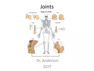
CLASSIFICATION • Joints are classified according to the tissues that lie between the bones into: • Fibrous. • Cartilaginous. • Synovial.
FIBROUS JOINTS • The articulating surfaces are joined by fibrous tissue. • Sutures of the vault of the skull: No movement, temporary joints (ossify later). • Inferior tibiofibularjoints (syndesmosis): Little movement, permanent joints.
CARTILAGINOUS JOINTS • Primary Cartilaginous • The bones are united by a plate or bar of hyaline cartilage. • No movement, temporary joints (ossify later). • Between the Epiphysis and Diaphysisof a growing bone. • Between the First Rib and the Sternum (1ststernocostal joint).
CARTILAGINOUS JOINTS • Secondary Cartilaginous • The bones are united by a plate of fibrocartilage. • Their articulating surfaces are covered by a thin plate of hyaline cartilage. • Little movement, permanent joints. • Midline joints. • Joints between the Vertebral Bodies(Intervertebral discs). • Symphysis Pubis.
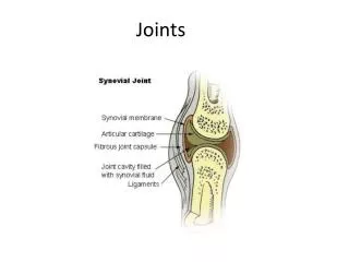
SYNOVIAL JOINTS • Characteristic features: • Freely movable joints. • A fibrous capsule attached to margins of articular surfaces & enclosing the joint. • The articular surfaces are covered by a thin layer of hyaline cartilage (articular cartilage). • A joint cavity enclosed within the capsule. Capsule Articular cartilage Articular cartilage
SYNOVIAL JOINTS • A thin vascular synovial membrane lining the inner surface of capsule. • A lubricating synovial fluid produced by synovial membrane in the joint cavity. It minimizes friction between articular surfaces. Synovial membrane Capsule containing synovial fluid
CLASSIFICATION OF SYNOVIAL JOINTS • Synovial joints are classified according to the range of movement into: • Plane synovial joints. • Axial synovial joints.
PLANE SYNOVIAL JOINTS • The articulating surfaces are flat and the bones slide on one another, producing a gliding movement. • Intercarpal Joints. • Sternoclavicular and Acromioclavicularjoints.
AXIAL SYNOVIAL JOINTS • Movements occur along axes: • Transverse: flexion & extension occur. • Longitudinal: rotation occurs. • Antero-posterior: abduction & adduction occur. • Axial joints are divided into: • Uniaxial. • Biaxial. • Multi-axial (polyaxial).
UNIAXIAL SYNOVIAL JOINTS • Hinge joints: • Axis: transverse. • Movements: flexion & extension. • Example: elbow joint. • Pivot: • Axis: longitudinal. • Movements: rotation. • Example:radio-ulnar joints
BIAXIAL SYNOVIAL JOINTS • Ellipsoid joints: • An elliptical convex fits into an elliptical concave articular surface. • Axes: Transverse & antero-posterior. • Movements: Flexion & extension + abduction & adduction. • Example: Wrist joint.
BIAXIAL SYNOVIAL JOINTS • Saddle joints: • The articular surfaces are reciprocally concavoconvex. • They resemble a saddle on a horse’s back. • Movement: As ellipsoid joints (Flexion & extension + abduction & adduction) + a small range of dependant rotation rotation. • Example:Carpometacarpal joint of the Thumb.
POLYAXIAL SYNOVIAL JOINTS • Ball-and-socket joints: • A ball –shaped head of one bone fits into a socket like concavity of another. • Movements: Flexion & extension + abduction & adduction) + rotation along a separate axis. • Examples: • Shoulder joint. • Hip Joint.
STABILITY OF SYNOVIAL JOINTS • The shape of articular surfaces: • The ball and socket shape of the Hip joint is a good examples of the importance of bone shape to maintain joint stability. • The shape of the bones forming the Knee joint has nothing to do for stability.
STABILITY OF SYNOVIAL JOINTS • The strength of ligaments: • They prevent excessive movement in a joint.
STABILITY OF SYNOVIAL JOINTS • The tone of the surrounding muscles: • In most joints, it is the major factor controlling stability. • The short muscles around the shoulder joint keeps the head of the humerus in the shallow glenoid cavity.
NERVE SUPPLY OF JOINTS • The capsule and ligaments receive an abundant sensory nerve supply. • Hilton’s Law: “A sensory nerve supplying a joint also supplies the muscles moving the joint and the skin overlying the insertions of these muscles.”
SUMMARY • Joint is the site where two or more bones come together, whether or not movement occurs between them. • Joints are classified according to the tissues that lie between the bones into: fibrous, cartilaginous & synovial. • Synovial joints are freely movable & characterized by the presence of : fibrous capsule, articular cartilage, synovial membrane & joint cavity containing synovial fluid.
SUMMARY • Synovial joints are classified according to the range of movement into: plane & axial. • Axial are divided according to the number of axes of movements into: uni-, bi- & polyaxial. • Stability of synovial joints depends on: shape of articular surfaces, ligaments & muscle tone. • Joints have same nerve supply as muscles moving them.
QUESTION 1 • In the synovial joint : • articular surfaces are united by a plate of fibrocartilage. • the synovial membrane is not vascular. • stability is not related to muscle tone. • movement is free.
QUESTION 2 • The elbow joint: • is a fibrous joint. • is a secondary cartilaginous joint. • allows only flexion & extension. • Is a synovial pivot joint.