Download
1 / 60
800 likes | 2.2k Views
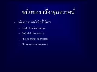
ชนิดของกล้องจุลทรรศน์. กล้องจุลทรรศน์ชนิดที่ใช้แสง Bright-field microscope Dark-field microscope Phase-contrast microscope Fluorescence microscopes. กล้องจุลทรรศน์แบบใช้แสง. กำลังขยายโดยทั่วไป 1000 – 2000 เท่า ภาพที่ได้จะเป็นภาพที่ทึบ พื้นหลังสว่าง ประกอบด้วยเลนส์ใกล้วัตถุหลายอัน

E N D
ชนิดของกล้องจุลทรรศน์ชนิดของกล้องจุลทรรศน์ • กล้องจุลทรรศน์ชนิดที่ใช้แสง • Bright-field microscope • Dark-field microscope • Phase-contrast microscope • Fluorescence microscopes
กล้องจุลทรรศน์แบบใช้แสงกล้องจุลทรรศน์แบบใช้แสง • กำลังขยายโดยทั่วไป 1000 – 2000 เท่า • ภาพที่ได้จะเป็นภาพที่ทึบ พื้นหลังสว่าง • ประกอบด้วยเลนส์ใกล้วัตถุหลายอัน • parfocal microscopes remain in focus when objectives are changed • กำลังขยายของภาพ = กำลังขยายของเลนส์ใกล้ตา x กำลังขยายของเลนส์ใกล้วัตถุ
รายละเอียดของภาพ (Resolution power) • ความสามารถในการแยกจุด 2 จุดออกจากกัน • ความยาวคลื่นแสง shorter wavelength greater resolution
Eyepiece กล้องจุลทรรศน์ Binocular tube Revolving nosepiece Arm Objective Mechanical stage Fine adjustment Stage Course adjustment Condenser Illuminator Base
ความสามารถในการขยายภาพของกล้องจุลทรรศน์ความสามารถในการขยายภาพของกล้องจุลทรรศน์ • Resolution power • ความสามารถของเลนส์ใกล้วัตถุในการแยกจุดสองจุดที่อยู่ใกล้กัน R = 0.6 x /NA • Numerical aperture • ความสามารถของเลนส์ใกล้วัตถุในการเก็บรวบรวมแสงที่หักเหภายในวัตถุ NA = n sin
Working distance Low power High power
Working distance Oil Immersion
Property Objective lens Scanning Low power High power Oil Immersion magnification 4x 10x 40-45x 90-100x Numerical aperture 0.1 0.25 0.55-0.65 1.25-1.4 Focal length 40 mm 16 mm 4 mm 1.8-2.0 mm Working distance 17-20 mm 4-8 mm 0.5-0.7 mm 0.1 mm Resolution power (WL= 450 nm; blue light) 2.3 m 0.9 m 0.35 m 0.18 m • คุณสมบัติของเลนส์ใกล้วัตถุ • working distance • - ระยะระหว่างเลนส์ใกล้วัตถุ กับผิวของแผ่น กระจกปิดตัวอย่าง หรือ ตัวอย่าง
The 3 Classes of Objectives Chromatic and Mono-Chromatic Corrections
กล้องพื้นหลังมืด (Dark-Field Microscope) • ภาพที่เกิดจะสว่างกว่าพื้นหลัง • ใช้ในการศึกษาตัวอย่างที่มีชีวิต และไม่ต้องการย้อมสี ns
Phase-contrast microscope • ภาพที่เกิดจะแสดงให้เห็นความแตกต่าง โครงสร้างภายในที่เกิดจากการหักเหของแสงที่แตกต่าง • ใช้ในการศึกษาเซลล์ที่มีชีวิต • โปร่งใสและไม่ต้องย้อมสี • เพิ่ม annual diaphragm ใต้ condenser และ phase plate ใน objective lens • wave length ~ 1/4 ของปกติ
Fluorescence Microscope • ใช้แสง ultraviolet, violet หรือ blue light • ตัวอย่างถูกย้อมด้วย สี fluorochromes • ภาพที่เกิด ตัวอย่างหรือ ส่วนที่ถูกย้อมด้วยสีจะเรืองแสง และพื้นหลังจะมืด
ภาพที่เกิดจากกล้อง Fluorescence Microscope
การเตรียมตัวอย่างและการย้อมสีการเตรียมตัวอย่างและการย้อมสี • เพิ่มความสามารถในการมองเห็นตัวอย่างให้ชัดเจนขึ้น • ช่วยให้เห็นรูปร่างลักษณะของตัวอย่างได้ชัดเจนขึ้น • เก็บรักษาตัวอย่าง
Fixation • หยุดกิจกรรมต่างๆ ภายในเซลล์ • เป็นขั้นตอนที่รักษาโครงสร้างทั้งภายนอกและภายในตัวอย่างให้อยู่ในสภาพคงที่ • เป็นการทำให้ตัวอย่างติดแน่นอยูบนแผ่นกระจกสไลด์ • โดยการใช้ความร้อน • เป็นการรักษารูปร่างให้คงที่แต่ไม่รักษาโครงสร้างภายใน • โดยการใช้สารเคมี • เป็นการรักษาโครงสร้างภายใน รูปร่าง เช่น Formaldehyde, Odmium tetroxide (Os4) , potassium permanganate, Glutaraldehyde
การทำให้แห้ง และย้อมสีแบบง่าย (dehydration and simple staining) • สี (stain) • ทำให้สามารถมองเห็นโครงสร้างภายในและภายนอกได้ชัดเจนยิ่งขึ้น และแตกต่างจากพื้นหลัง • การย้อมสีแบบง่าย • ใช้สีเพียงสีเดียว • เช่น สีเบสิค (basic dyes)
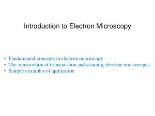
Electron Microscopy • ใช้ลำแสงอิเล็คตรอนในการสร้างภาพ • ความยาวคลื่นแสงสั้นกว่าแสงทำให้ภาพที่เกิดขึ้นมีรายละเอียดสูงขึ้น
Transmission Electron Microscope • electrons scatter when they pass through thin sections of a specimen • transmitted electrons (those that do not scatter) are used to produce image • denser regions in specimen, scatter more electrons and appear darker
The Scanning Electron Microscope • uses electrons reflected from the surface of a specimen to create image • produces a 3-dimensional image of specimen’s surface features
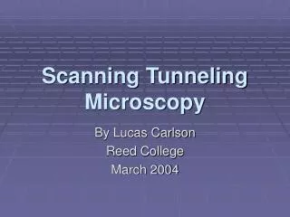
Confocal microscopy • have extremely high resolution • can be used to observe individual atoms
Confocal Microscopy • confocal scanning laser microscope • laser beam used to illuminate spots on specimen • computer compiles images created from each point to generate a 3-dimensional image
Biochemical Techniques วัตถุประสงค์ เพื่อศึกษาคุณสมบัติและหน้าที่ของส่วนประกอบต่างๆ ของเซลล์ Molecule Small Assembly of macromolecule Large
หลักการ คือ การทำให้เซลล์แตกเป็นชิ้นส่วน (Cell fractination) และเก็บชิ้นส่วนของเซลล์มาศึกษา โดย 1. การทำให้เป็นเนื้อเดียวกัน (Homogenization) สามารถทำได้หลายวิธีคือ 1. โดยการบด (Pestle /Moltar) 2. การใช้คลื่นเสียง (Ultrasonic vibration) 3. การสับเป็นชิ้นๆ (Wareing bleden/ Potter homogenizers) 4. การทำให้แข็งตัวและละลาย (Freeze and Thaw) 5. การอัดด้วยความดันสูง (Deactivation) 6. การใช้สารเคมี (Chemical) 7. การใช้เอนไซม์ (Enzyme)
Homogenizer http://www.freewebs.com/ltaing/chpt7.3Cellfractionation.gif
Gel Filtration http://fig.cox.miami.edu/~cmallery/150/protein/proteinsb.htm
2. การแยกส่วนประกอบต่างๆ ออกจากกัน (Separation of the homogenate) 1. การปั่นตกตะกอน (Centrifugation) - Gravity (g) - Relative centrifugal force (RFC) - Rate of sedimentation of components (size, shape, density of particle and solvent, speed) - Differential centrifugation - Rate-zonal or density-gradient centrifugation
Differential Centrifigation http://www.freewebs.com/ltaing/chpt7.3Cellfractionation.gif
Differential Centrifigation http://www.freewebs.com/ltaing/chpt7.3Cellfractionation.gif
3. การแยกชิ้นส่วนของเซลล์ (Separation of biomolecules) 1. ความสามารถในการละลาย (Solubility) 2. ขนาด (size) - Centrifugation - Dialysis - Ultrafiltration - Gel filtration - SDS -PAGE (Sodium Dodecyl Sulphate Polyacrylamide Gel Electrophoresis)
3. ประจุไฟฟ้า (Charge) Ion exchange chromatography Isoelectric focusing Electrophoresis 4. Biological properties Affinity of chromatography Immunoelectrophoresis Blotting - Southern blot (DNA) - Northern blot (RNA) - Western blot (Protein) Chromatography อื่นๆ (paper. Gas , Thin-layer) PCR (Polymerase chain reaction)
Ion Exchange http://fig.cox.miami.edu/~cmallery/150/protein/ion_exchange.jpg