Download
1 / 29
290 likes | 598 Views
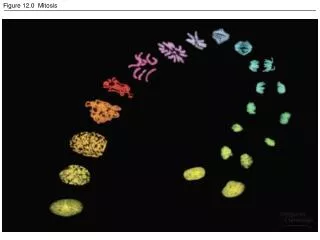
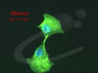
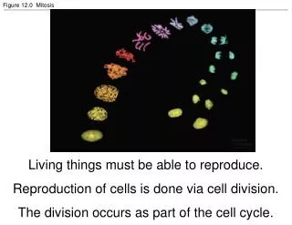
Figure 12.0 Mitosis. Living things must be able to reproduce. Reproduction of cells is done via cell division. The division occurs as part of the cell cycle. Figure 12.1a The functions of cell division: Reproduction. Purposes: Reproduction of cells Repair of tissue Growth Pass on DNA.

E N D
Figure 12.0 Mitosis Living things must be able to reproduce. Reproduction of cells is done via cell division. The division occurs as part of the cell cycle.
Figure 12.1a The functions of cell division: Reproduction Purposes: Reproduction of cells Repair of tissue Growth Pass on DNA
Figure 12.1b The functions of cell division: Growth and development Cell division is also central to the development of a multicellular organism that begins as a fertilized egg or zygote.
Figure 12.1c The functions of cell division: Tissue renewal • Multicellular organisms also use cell division to repair and renew cells that die from normal wear and tear or accidents. • A cell’s genetic information, packaged as DNA, is called its genome. • In prokaryotes, the genome is often a single long DNA molecule. • In eukaryotes, the genome consists of several DNA molecules.
Figure 12.2 Eukaryotic chomosomes • DNA molecules are packaged into chromosomes. • Every eukaryotic species has a characteristic number of chromosomes in the nucleus. • Human somaticcells (body cells) =46 chromosomes. • Human gametes (sperm or eggs) = 23 chromosomes • Fertilization fuses two gametes together and doubles the number of chromosomes to 46 again.
Figure 12.3 Chromosome duplication and distribution during mitosis • Chromosomes have a DNA and protein complex, chromatin, organized into a long thin fiber.
Figure 12.4 The cell cycle Interphase: [90%] G1 (growth 1), S (synthesis) and G2 (growth 2) M Phase [10%] – mitosis and cytokinesis
Figure 12.5 The stages of mitotic cell division in an animal cell: G2 phase; prophase; prometaphase • chromosomes are tightly coiled • The nucleoli disappear. • The mitotic spindle begins to form • cell completes preparations for cell division. • the nuclear envelope fragments and microtubules from the spindle interact with the chromosomes. • Microtubules from one poles attach the kinetochores,
Figure 12.5 The stages of mitotic cell division in an animal cell: metaphase; anaphase; telophase and cytokinesis. • The cell continues to elongate • Two nuclei begin to form • Cytokinesis, division of the cytoplasm, begins. • At anaphase, the centromeres divide, separating the sister chromatids. (Pulled toward the poles) • The spindle fibers push the sister chromatids until they are all arranged at the metaphase plate,
Assembly of the spindle microtubules starts in the centrosome. The centrosome (microtubule-organizing center) of animals has a pair of centrioles at the center, but the function of the centrioles is somewhat undefined. Figure 12.6 The mitotic spindle at metaphase
One hypothesis for the movement of chromosomes in anaphase is that motor proteins at the kinetochore “walk” the attached chromosome along the microtubule toward the opposite pole. • The excess microtubule sections depolymerize. • Experiments support the hypothesis that spindle fibers shorten during anaphase from the end attached to the chromosome, not the centrosome. • Nonkinetichore microtubules are responsible for lengthening the cell along the axis defined by the poles. Figure 12.7 Testing a hypothesis for chromosome migration during anaphase
Figure 12.10 Bacterial cell division (binary fission) (Layer 1)
Figure 12.10 Bacterial cell division (binary fission) (Layer 2)
Figure 12.10 Bacterial cell division (binary fission) (Layer 3)
Figure 12.12 Evidence for cytoplasmic chemical signals in cell cycle regulation • The cell cycle appears to be driven by specific chemical signals in the cytoplasm. • Fusion of an S phase cell and a G1 phase cell induces the G1 nucleus to start S phase. • Fusion of a cell in mitosis with one in interphase induces the second cell to enter mitosis.
The distinct events of the cell cycle are directed by a distinct cell cycle control system. • These molecules trigger and coordinate key events in the cell cycle. • The control cycle has a built-in clock, but it is also regulated by external adjustments and internal controls • For many cells, the G1 checkpoint, the restriction point in mammalian cells, is the most important.(go ahead or G0 phase. Figure 12.13 Mechanical analogy for the cell cycle control system
Figure 12.14 Molecular control of the cell cycle at the G2 checkpoint Peaks in the activity of one cyclin-Cdk complex, MPF, correspond to peaks in cyclin concentration. • MPF (“maturation-promoting factor” or “M-phase-promoting-factor”) triggers the cell’s passage past the G2 checkpoint to the M phase. • MPF promotes mitosis by phosphorylating a variety of other protein kinases. • MPF stimulates fragmentation of the nuclear envelope. • It also triggers the breakdown of cyclin, dropping cyclin and MPF levels during mitosis and inactivating MPF
A variety of external chemical and physical factors can influence cell division. Particularly important for mammalian cells are growth factors, proteins released by one group of cells that stimulate other cells to divide. For example, platelet-derived growth factors (PDGF), produced by platelet blood cells, bind to tyrosine-kinase receptors of fibroblasts, a type of connective tissue cell. This triggers a signal-transduction pathway that leads to cell division. Each cell type probably responds specifically to a certain growth factor or combination of factors.
The role of PDGF is easily seen in cell culture. • Fibroblasts in culture will only divide in the presence of a medium that also contains PDGF. • In a living organism, platelets release PDGF in the vicinity of an injury. • The resulting proliferation of fibroblasts helps heal the wound. Figure 12.15 The effect of a growth factor on cell division
Figure 12.16 Density-dependent inhibition of cell division • Growth factors appear to be important in density-dependent inhibition of cell division.
Figure 12.17 The growth and metastasis of a malignant breast tumor • If the abnormal cells remain at the originating site, the lump is called a benign tumor. • Most do not cause serious problems and can be removed by surgery. • In a malignant tumor, the cells leave the original site to impair the functions of one or more organs. • This typically fits the colloquial definition of cancer. • In addition to chromosomal and metabolic abnormalities, cancer cells often lose attachment to nearby cells, are carried by the blood and lymph system to other tissues, and start more tumors in a event called metastasis.
Figure 12-17x2 Mammogram: normal (left) and cancerous (right)