Download
1 / 18
210 likes | 632 Views
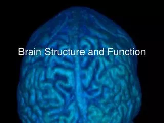
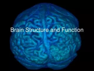
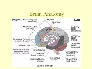
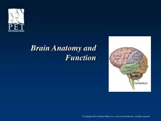
Brain Anatomy and Function. Anatomy of the Brain. Separated into right and left halves by the Interhemispheric Fissure. The Central Sulcus runs down & forward The Lateral Fissure runs backward & up. Frontal and Temporal Lobes. Thought Voluntary movement Speech motor

E N D
Anatomy of the Brain • Separated into right and left halves by the Interhemispheric Fissure • The Central Sulcus • runs down & forward • The Lateral Fissure runs backward & up
Frontal and Temporal Lobes • Thought • Voluntary movement • Speech motor • Covers 1/3rd of area of the brain Frontal Temporal • Memory • Auditory function
Parietal and Occipital Lobes • Sensation • Touch • Pressure • Pain • Temperature • Texture • Position/spatial orientation Parietal Occipital • Vision • Visual processes • Reading
Medulla Oblongata, Cerebellum, and Pons • Large Muscle Coordination • Balance • Walking, Writing Medulla Oblongata Cerebellum • Respiration • Heart rate • Continuous with the spinal cord (2.5 cm) Pons • Relay between the cerebral hemispheres and the cerebellum
Basal Ganglia and Thalamus “The Brakes” • Modifies movement on a minute-to-minute basis • Inhibits Movement • Coordination • Cortical relay
Limbic System • Attention • Sensory gateway • Memory processing • Rage • Aggression • Sexuality • Appetite/Thirst
The Nerve Cell Synaptic junction
Neurotransmitters • Serotonin – major – emotions, judgment, eating and sleep disorders (associated with frontotemporal disorder) • Glutamate/GABA - Widespread, anxiety, sleep, (Valium targets this) • Dopamine – memory, mood, movement, Parkinson's Disease, psychiatric problems • Endorphins – relief of pain, (Morphine targets this) Lichtman, J., et al Washington University 2002
Normal functions Emotions Judgment Sleep Imbalances Depression Suicidal behavior Anxiety Impulsive behavior Eating disorders Serotonin Glutamate/GABA • Normal functions • Involved in most facets of brain function • Imbalances • Memory disturbances • Sleep disturbances • Anxiety
Normal functions Mood Movement Memory Imbalances Movement disorders Schizophrenia Addiction Dopamine Endorphins • Normal functions • Relieve pain • Induce euphoria
Normal Aging Brain • Brain weight and volume decrease • Grooves widen • Surface smoothes • Neurofibrillary tangles increase • Understanding normal variation is key to interpretation
Brain Glucose Metabolism –Normal • Normal brain tissue actively metabolizes glucose and its analogue (F-18 FDG) • Glucose metabolism provides 95% of the energy required for brain function • FDG is irreversibly trapped within brain cells in proportion to its use because it cannot be broken down or stored unlike glucose
FDG-PET Abnormal Brain Imaging • Dementia • Memory loss • Cognitive Decline • Epilepsy • Localization of a seizure focus • Tumor Assessment • Radiation Necrosis vs Tumor • Grade • Objective Imaging Diagnosis of Movement Disorders • Huntington’s Disease • Parkinson’s Disease
Dementia Diagnosis:Current Methods • History and physical examination • Neurologist (Sens. = 50-80%) • Neuropsychologist / Neuropsychiatrist • Neuropsychological testing • MRI / CT • Blood testing • Functional Neuroimaging (SPECT/ PET/MR) • Sens.=80-90%
Summary • Normal Brain Anatomy • Normal Brain Function • Current PET Brain Applications: • Diagnosis of Dementia • Seizure Localization • Tumor Assessment • Objective Imaging Diagnosis of Movement Disorders (not CMS approved)
Contributors • Rebecca Trunnell Hyman • Coordinator of PET Services • Clinical PET of West County - Creve Coeur, MO • Kevin L. Berger, M.D. • Assistant Professor of Radiology • Director of PET Imaging • Michigan State University – East Lansing, MI