Download
1 / 36
370 likes | 615 Views
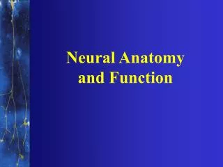
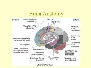
Overview of Brain Anatomy and function . Wei-Ching Lee, M.D. INTRODUCTION. Lobes Frontal Parietal Temporal Occipital Brainstem. Anatomy. Anatomy. Homunculus Man. Circle of Willis. Gold: ACA Pink: MCA Blue: PCA. Frontal Lobe. Conscientiousness Judgments

E N D
Overview of Brain Anatomy and function Wei-Ching Lee, M.D.
INTRODUCTION • Lobes • Frontal • Parietal • Temporal • Occipital • Brainstem
Frontal Lobe • Conscientiousness • Judgments • How we initiate activity in response to our environment. • Controls our emotional response. • Controls our expressive language. • Assigns meaning to the words we choose (abstract thought) • Attention span • Involves word associations (language planning) • Memory for habits and motor activities (short term memory) • Motor cortex—Voluntary movement • Impulse control • Perseverance
Frontal Lobe Deficit—Problems • Loss of simple movement of various body parts (Paralysis). • Inability to plan a sequence of complex movements needed to complete multi-stepped tasks, such as making coffee (Sequencing). • Loss of spontaneity in interacting with others. • Loss of flexibility in thinking. • Persistence of a single thought (Perseveration). • Inability to focus on task (Attending). • Mood changes (Emotionally Labile). • Changes in social behavior. • Changes in personality. • Difficulty with problem solving. • Inablility to express language (Broca's Aphasia).
Parietal Lobe Function • Location for visual attention. • Location for touch perception. • Goal directed voluntary movements. • Manipulation of objects. • Integration of different senses that allows for understanding a single concept.
Parietal Lobe—Problems resulting from deficit • Inability to attend to more than one object at a time. • Inability to name an object (Anomia). • Inability to locate the words for writing (Agraphia). • Problems with reading (Alexia). • Difficulty with drawing objects. • Difficulty in distinguishing left from right. • Difficulty with doing mathematics (Dyscalculia). • Lack of awareness of certain body parts and/or surrounding space (Apraxia) that leads to difficulties in self-care. • Inability to focus visual attention. • Difficulties with eye and hand coordination.
Temporal Lobe Function • Hearing ability • Memory acquisition • Some visual perceptions • Categorization of objects.
Temporal Lobe Deficits—Problems • Difficulty in recognizing faces (Prosopagnosia). • Difficulty in understanding spoken words (Wernicke's Aphasia). • Disturbance with selective attention to what we see and hear. • Difficulty with identification of, and verbalization about objects. • Short-term memory loss. • Interference with long-term memory • Increased or decreased interest in sexual behavior. • Inability to catagorize objects (Catagorization). • Right lobe damage can cause persistant talking. • Increased aggressive behavior.
Occipital Lobe Function • Vision
Occipital Lobe Deficits--Problems • Defects in vision (Visual Field Cuts). • Difficulty with locating objects in environment. • Difficulty with identifying colors (Color Agnosia). • Production of hallucinations • Visual illusions - inaccurately seeing objects. • Word blindness - inability to recognize words. • Difficulty in recognizing drawn objects. • Inability to recognize movement of an object (Movement Agnosia). • Difficulties with reading and writing.
Cerebellum Function • Coordination of voluntary movement • Balance and equilibrium • Some memory for reflex motor acts.
Cerebellum Deficits—Problems • Loss of ability to coordinate fine movements. • Loss of ability to walk. • Inability to reach out and grab objects. • Tremors. • Dizziness (Vertigo). • Slurred Speech (Scanning Speech). • Inability to make rapid movements.
Brainstem • Midbrain • Pons • Medulla
Brainstem Function • Breathing • Heart Rate • Swallowing • Reflexes to seeing and hearing (Startle Response). • Controls sweating, blood pressure, digestion, temperature (Autonomic Nervous System). • Affects level of alertness. • Ability to sleep. • Sense of balance (Vestibular Function).
Brainstem Deficits—Problems • Decreased vital capacity in breathing, important for speech. • Swallowing food and water (Dysphagia). • Difficulty with organization/perception of the environment. • Problems with balance and movement. • Dizziness and nausea (Vertigo). • Sleeping difficulties (Insomnia, sleep apnea).
Midbrain Function: • Body posture • Equilibrium • Autonomic Nervous System • Blood pressure • Temperature • Emotional influence • Reg appetite and hormones • Nuclei of CN III and IV
Midbrain lesion • Variable LOC • Abnormal extensor tone • Hyperventilation • CN III and IV deficits • CN IV nerve lesion: head tilted away from lesion • CN IV nucleus lesion: head tiled towards lesion • CN III: innervates all eyes muscles except LR6 and SO4, eye deviated laterally and downward with eyelid down (levator palpebrae)
Pons • Function • Respiration • Chewing • Taste • Arousal, wakefulness, alertness • Nuclei of CN V, VI, VII, VIII
Pons lesion • Semi-coma • Abnormal extensor tone • Apneusis • Withdrawal • CN V,VI, VII (facial colliculus syndrome) • CN V: ipsi jaw deviation upon opening • VI: diplopia, paralysis of ipsi LR but also inablity to turn contra eye medially • VII: can’t close eye or smile
Medulla Function: • Life-sustaining control center: controls hear, respiration, vasomotor • Cough, gag, swallow, vomit, digest • Nuclei of CN VIII, IX, X, XI, XII
Medulla Lesion • Comatose • Abnormal breathing • Ataxic • Absent gag reflex • Absent cough • CN VIII, IX, X, XI, XII deficits • VIII: ipsi stumbling but contra nystagmus • IX, X, XI: absent gag reflex, contra uvula deviation, dysphonia, dysphagia • XII: ipsi tongue deviation and atrophy
Function of Hemispheres Right Hemisphere • judging the position of things in space • knowing body position • understanding and remembering things we do and see • putting bits of information together to make an entire picture • controls the left side of the body Left Hemisphere • understanding and use of language (listening, reading, speaking and writing) • memory for spoken and written messages • detailed analysis of information • controls the right side of the body
Online references • http://www.wisc-online.com/objects/index_tj.asp?objid=OTA502 • http://www.neuroskills.com/edu/ceufunction1.shtml • http://www.hopkinshospital.org/health_info/Neurological%20Diseases/Reading/brain_anatomy.html • http://training.seer.cancer.gov/ss_module00_bbt/unit02_sec04_c_brain.html
SAE • Findings commonly seen after right hemisphere stroke include • Right hemiplegia • Aphasia • Visual-Perceptual deficits • Agraphia
SAE • Answer C • Strokes on nondominant hemisphere present with contralateral hemiplegia and hemianesthesia, aprosody (absence of normal speech in pitch, rhythm, and variations in stress), visual spatial deficit, and neglect syndrome.
SAE • In TBI, MRI is preferred to CT scan in the • Eval of acute brain injury • Detection of SAH • Detection of epidural hematomas • Eval of diffuse axonal injury
SAE • Answer D • MRI is considered better than CT for evaluating DAI. CT is superior to MRI for detection of acute extra-axial hematomas, and in the eval of acute brain injury
SAE • 74 y/o woman has had a stroke with left hemiparesis and left neglect. Muscle tone is increased, and flexion contractures are beginning to develop in her left elbow, wrist, and hand. Initial intervention would be • Diazepam 2.5mg tid • Neurolytic block to median nerve • Botulinum toxin injection to forearm flexors • Static muscle stretch • Baclofen 5mg qid
SAE • Answer D. • In treating spasticity, the approach with the least possible adverse effects should be used first. In this case, ROM, stretching, and positioning with splints would be the initial treatment.
SAE • Following a head injury, a 35 y/o W presents with vertigo. She reports a sensation of spinning beginning several seconds after standing up radiply, bending over, or rolling in bed. Symptoms lasts for approx 30 sec. Exam is notable for nystagmus during episodes of vertigo, normal extremity coordination, and min increase in sway during Romberg. Most likely dx is: • Benign positional vertigo • Cerebellar contusion • Unilateral vestibular paresis • Bilateral vestibular paresis
SAE • Answer A. • BPV characterized by transient episodes of vertigo precipitated by changes in position of the head. Treatment involves psecific otolith repositioning maneuver or seris of habituation exercises.