Download
1 / 27
290 likes | 396 Views
Agarose gel electrophoresis is a method to separate and analyze DNA fragments based on size and charge. Learn about components, concentrations, and running the gel effectively for DNA analysis. Visualization with ethidium bromide under UV light helps in identifying DNA fragments.

E N D
Agarose Gel Electrophoresis
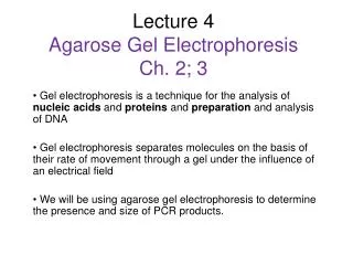
Gel electrophoresisis a method for separation and analysis of macromolecules(DNA, RNA and proteins) and their fragments, based on their size and charge.
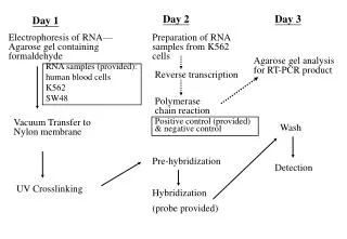
Applications of Agarose Gel Electrophoresis • Separation of restriction enzyme digested DNA including genomic DNA. • Analysis of PCR products after polymerase chain reaction to assess for target DNA amplification. • Allows for the estimation of the size of DNA molecules using a DNA marker or ladder which contains DNA fragments of various known sizes. • Allows the rough estimation of DNA quantity and quality.
Components of an Electrophoresis System • Gel: a porous material that molecules migrates through: * Gel can be made from substances such as agarose or polyacrylamide.
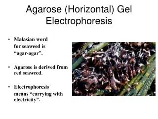
Agarose “ a complex sugar chain from red seaweed”. *Non toxic carbohydrate. *It is commonly used in foods (ice cream, and jellies) and many biological mediums. *It has a large pore size good for separating large molecules quickly. • *Acts as a sieve for separating molecules. • *This solid matrix will allow the separation of fragments by size.
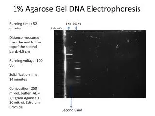
Concentration of the gel affects molecules migration :- *Most agarose gels are made with between 0.7% (good separation or resolution of large 5–10kb DNA fragments) and 2% (good resolution for small 0.2–1kb fragments). *Up to 3% can be used for separating very tiny fragments. *Low percentage gels are very weak and may break when you try to lift them. *1% gels are common for many applications. *Agarose gels do not have a uniform pore size.
1% agarose 2% agarose • Low conc. = larger pores better resolution of larger DNA fragments • High conc. = smaller pores better resolution of smaller DNA fragments • So smaller molecules move faster and migrate farther than larger ones because smaller molecules migrate more easily through the pores of the gel. • This phenomenon is called sieving.
Porous Material Molecules Entering Porous Material Smallest Move Fastest Molecule Size
2. Power supply and chamber: a source of power supply * During electrophoresis, water is electrolyzed which generates protons (H+ ions) at the anode (positive) and hydroxyl ions (OH -1) at the cathode (negative). The cathode (negative) end of the electrophoresis chamber then becomes basic and the anode (positive) end becomes acidic. * The electrode at which electrons enter the gel box from the power supply (along the black wire) is called the cathode . The electrode at which electrons leave the box and re-enter the power supply (along the red wire) is called the anode and . Power Supply
*The flow of electrons sets up a potential energy difference between the electrodes. This is known as potential, and is measured in volts.
Cathode - Anode + Buffer Dyes
So molecules with a negative charge (anions) will be attracted to the positively charged node (anode)……. Red color. Molecules with a positive charge (cations) will be attracted to the negatively charged node (cathode)……Black color. DNA has negative charge which migrate from cathode to anode
Positive Molecules Analyze Identify Purify Size Separation Charge Separation Mixture of Charged Molecules Negative Molecules Separation of a Mixture of Charged Molecules Charged molecules are separated based on their electrical charge and size.
3. Buffer: a fluid mixture of water and ions. *A buffer is a chemical system that maintains a relatively constant pH even when strong acids or bases are added. Buffer solutions contain either a weak acid or weak base and one of their salts. Because a change in pH can alter the charge on a particle, it is important to use a buffer solution when separating during electrophoresis.
Electrophoresis Buffer • TAE (Tris -acetate-EDTA) and TBE (Tris-borate-EDTA) – pH buffer. • Tris a pH buffer. • Acetic acid provide ions to support conductivity and maintain pH. • EDTA, prevent brake down of molecules. “all dissolved in water”. Notes: Use of water will produce no migraton Agarose dissolved in electrophoresis buffer
Overview of Agarose Gel Electrophoresis • Gel Preparation • Loading the gel • Running the gel
Gel Preparation Agarose is a linear polymer extracted from seaweed.
Agarose Buffer Solution Combine the agarose powder and buffer solution. Use a flask that is several times larger than the volume of buffer.
Melting the Agarose Agarose is insoluble at room temperature (left). The agarose solution is boiled until clear (right).
Gels are covered with a buffer solution. Prior to loading the samples. • The DNA must be mixed with a loading dye. • The loading dye serves two purposes: • Increases the density of the DNA so it will sink into the wells. • Provides a visual marker so you know how far the DNA (which is not visible) has traveled in the gel.
Visualization • DNA may be visualized using ethidium bromide which, when intercalated into DNA, fluoresce under ultraviolet light.