Download
1 / 55
560 likes | 741 Views
Learn about agarose gel electrophoresis, a technique to separate DNA molecules by size and charge, used in DNA analysis. Follow step-by-step instructions for preparing and running a gel.

E N D
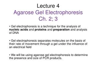
Agarose Gel Electrophoresis • Gel electrophoresis is a widely used technique for the analysis of nucleic acids and proteins. Agarose gel electrophoresis is routinely used for the preparation and analysis of DNA. • Gel electrophoresis is a procedure that separates molecules on the basis of their rate of movement through a gel under the influence of an electrical field. • We will be using agarose gel electrophoresis to determine the presence and size of PCR products. PCR products indicate the presence of Wolbachia.
How does electrophoresis work? • The gel is made from agar • DNA is a negative molecules • Molecules sort based on • Charge • Size • shape
Additional Information on Gel Electrophoresis: Virtual Gel Electrophoresis http://gslc.genetics.utah.edu/units/biotech/gel/
Agarose gel electrophoresis is a method to separate DNA, or RNA molecules by size. This is achieved by moving negatively charged nucleic acid molecules through an agarose matrix with an electric field (electrophoresis). Shorter molecules move faster and migrate farther than longer ones .
Analysis of PCR products, e.g. in molecular genetic diagnosis or genetic fingerprinting
• When placed in an electrical field, DNA will migrate toward the positive pole (anode). H O2 DNA - + Power • Polymerized agarose is porous, allowing for the movement of DNA Scanning Electron Micrograph of Agarose Gel (1×1 µm) • DNA is negatively charged. • An agarose gel is used to slow the movement of DNA and separate by size.
DNA - + Power How fast will the DNA migrate? strength of the electrical field, buffer, density of agarose gel… Size of the DNA! *Small DNA move faster than large DNA …gel electrophoresis separates DNA according to size small large Within an agarose gel, linear DNA migrate inversely proportional to the log10 of their molecular weight.
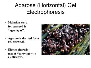
What is agar? Agar comes from sea weed. What is it used for? The gel is 1% agarous and has no electrical charge.
Agarose D-galactose 3,6-anhydro L-galactose • Sweetened agarose gels have been eaten in the Far East since the 17th century. • Agarose was first used in biology when Robert Koch* used it as a culture medium for Tuberculosis bacteria in 1882 *Lina Hesse, technician and illustrator for a colleague of Koch was the first to suggest agar for use in culturing bacteria Agarose is a linear polymer extracted from seaweed.
An agarose gel is prepared by combining agarose powder and a buffer solution. Buffer Flask for boiling Agarose
Electrophoresis Equipment Power supply Cover Gel tank Electrical leads Casting tray Gel combs
Preparing the Casting Tray Seal the edges of the casting tray and put in the combs. Place the casting tray on a level surface. None of the gel combs should be touching the surface of the casting tray.
Agarose Buffer Solution Combine the agarose powder and buffer solution. Use a flask that is several times larger than the volume of buffer.
Melting the Agarose Agarose is insoluble at room temperature (left). The agarose solution is boiled until clear (right). Gently swirl the solution periodically when heating to allow all the grains of agarose to dissolve. ***Be careful when boiling - the agarose solution may become superheated and may boil violently if it has been heated too long in a microwave oven.
Pouring the gel Allow the agarose solution to cool slightly (~60ºC) and then carefully pour the melted agarose solution into the casting tray. Avoid air bubbles.
Each of the gel combs should be submerged in the melted agarose solution.
When cooled, the agarose polymerizes, forming a flexible gel. It should appear lighter in color when completely cooled (30-45 minutes). Carefully remove the combs and tape.
DNA buffer wells Anode (positive) Cathode (negative) Add enough electrophoresis buffer to cover the gel to a depth of at least 1 mm. Make sure each well is filled with buffer.
Sample Preparation Mix the samples of DNA with the 6X sample loading buffer (w/ tracking dye). This allows the samples to be seen when loading onto the gel, and increases the density of the samples, causing them to sink into the gel wells. 6X Loading Buffer: Bromophenol Blue (for color) Glycerol (for weight)
Loading the Gel Carefully place the pipette tip over a well and gently expel the sample. The sample should sink into the well. Be careful not to puncture the gel with the pipette tip.
Running the Gel Place the cover on the electrophoresis chamber, connecting the electrical leads. Connect the electrical leads to the power supply. Be sure the leads are attached correctly - DNA migrates toward the anode (red). When the power is turned on, bubbles should form on the electrodes in the electrophoresis chamber.
Cathode (-) wells Bromophenol Blue DNA (-) Gel Anode (+) After the current is applied, make sure the Gel is running in the correct direction. Bromophenol blue will run in the same direction as the DNA.
12,000 bp 5,000 DNA migration 2,000 1,650 1,000 850 650 500 400 300 200 100 DNA Ladder Standard - Note: bromophenol blue migrates at approximately the same rate as a 300 bp DNA molecule bromophenol blue + Inclusion of a DNA ladder (DNAs of know sizes) on the gel makes it easy to determine the sizes of unknown DNAs.
Staining the Gel • Ethidium bromide binds to DNA and fluoresces under UV light, allowing the visualization of DNA on a Gel. • Ethidium bromide can be added to the gel and/or running buffer before the gel is run or the gel can be stained after it has run. ***CAUTION! Ethidium bromide is a powerful mutagen and is moderately toxic. Gloves should be worn at all times.
Staining the Gel • Place the gel in the staining tray containing warm diluted stain. • Allow the gel to stain for 25-30 minutes. • To remove excess stain, allow the gel to destain in water. • Replace water several times for efficient destain.
Ethidium Bromide requires an ultraviolet light source to visualize
DNA ladder DNA ladder 1 2 3 4 5 6 7 8 wells • 5,000 bp 2,000 1,650 1,000 850 650 500 400 PCR Product 300 200 100 + - - + - + + - Visualizing the DNA (ethidium bromide) Primer dimers Samples # 1, 4, 6 & 7 were positive for Wolbachia DNA
Visualizing the DNA (QuikVIEW stain) DNA ladder wells 2,000 bp PCR Product 1,500 1,000 750 500 250 + - - - - + + - - + - + Samples # 1, 6, 7, 10 & 12 were positive for Wolbachia DNA March 12, 2006
Visualisation: Ethidium Bromide (EtBr) and dyes • The most common dye used to make DNA or RNA bands visible for agarose gel electrophoresis is ethidium bromide, usually abbreviated as EtBr. It fluoresces under UV light when intercalated into DNA (or RNA). By running DNA through an EtBr-treated gel and visualizing it with UV light. EtBr is a known mutagen, however, safer alternatives are available.
Biotechnology Gel Electrophoresis
Many uses of restriction enzymes… • Now that we can cut DNA with restriction enzymes… • we can cut up DNA from different people… or different organisms… and compare it • why? • forensics • medical diagnostics • paternity • evolutionary relationships • and more…
Comparing cut up DNA • How do we compare DNA fragments? • separate fragments by size • How do we separate DNA fragments? • run it through a gelatin • gel electrophoresis • How does a gel work?
Gel electrophoresis • A method of separating DNA in a gelatin-like material using an electrical field • DNA is negatively charged • when it’s in an electrical field it moves toward the positive side DNA – + “swimming through Jello”
Gel electrophoresis • DNA moves in an electrical field… • so how does that help you compare DNA fragments? • size of DNA fragment affects how far it travels • small pieces travel farther • large pieces travel slower & lag behind DNA – + “swimming through Jello”
Gel Electrophoresis DNA &restriction enzyme - longer fragments wells power source gel shorter fragments completed gel +
Running a gel fragments of DNAseparate out based on size cut DNA with restriction enzymes Stain DNA • ethidium bromide binds to DNA • fluoresces under UV light 1 2 3
DNA fingerprint • Why is each person’s DNA pattern different? • sections of “junk” DNA • doesn’t code for proteins • made up of repeated patterns • CAT, GCC, and others • each person may have different number of repeats • many sites on our 23 chromosomes with different repeat patterns GCTTGTAACGGCCTCATCATCATTCGCCGGCCTACGCTT CGAACATTGCCGGAGTAGTAGTAAGCGGCCGGATGCGAA GCTTGTAACGGCATCATCATCATCATCATCCGGCCTACGCTT CGAACATTGCCGTAGTAGTAGTAGTAGTAGGCCGGATGCGAA
Allele 1 cut sites repeats cut sites GCTTGTAACGGCCTCATCATCATTCGCCGGCCTACGCTT CGAACATTGCCGGAGTAGTAGTAAGCGGCCGGATGCGAA GCTTGTAACG GCCTCATCATCATCGCCG GCCTACGCTT CGAACATTGCCG GAGTAGTAGTAGCGGCCG GATGCGAA DNA patterns for DNA fingerprints Cut the DNA 1 2 3 – + DNA allele 1
Differences between people Person 1 cut sites cut sites GCTTGTAACGGCCTCATCATCATTCGCCGGCCTACGCTT CGAACATTGCCGGAGTAGTAGTAAGCGGCCGGATGCGAA Person 2: more repeats GCTTGTAACGGCCTCATCATCATCATCATCATCCGGCCTACGCTT CGAACATTGCCGGAGTAGTAGTAGTAGTAGTAGGCCGGATGCGAA 1 2 3 DNA fingerprint – + DNA person 1 person 2
1 2 3 4 5 1 2 3 4 5 Uses: Evolutionary relationships • Comparing DNA samples from different organisms to measure evolutionary relationships turtle snake rat squirrel fruitfly – DNA +
Uses: Medical diagnostic • Comparing normal allele to disease allele chromosomewith normal allele 1 chromosome with disease-causing allele 2 allele 2 allele 1 – DNA Example: test for Huntington’s disease +
Uses: Forensics • Comparing DNA sample from crime scene with suspects & victim suspects crime scene sample S1 S2 S3 V – DNA +
DNA fingerprints • Comparing blood samples on defendant’s clothing to determine if it belongs to victim • DNA fingerprinting
RFLP / electrophoresis use in forensics • 1st case successfully using DNA evidence • 1987 rape case convicting Tommie Lee Andrews “standard” semen sample from rapist blood sample from suspect “standard” How can you compare DNA fromblood & from semen?RBC? “standard” semen sample from rapist blood sample from suspect “standard”
Electrophoresis use in forensics • Evidence from murder trial • Do you think suspect is guilty? blood sample 1 from crime scene blood sample 2 from crime scene blood sample 3 from crime scene “standard” blood sample from suspect OJ Simpson blood sample from victim 1 N Brown blood sample from victim 2 R Goldman “standard”
Mom F1 F2 child Uses: Paternity • Who’s the father? – DNA +
I’m a-glow! Got any Questions?
Biodiversity Lab Which plant is most closely related to Botanus curus?