Download
1 / 12
120 likes | 216 Views
Learn how Drosophila establishes its body plan through intricate gene expression patterns during embryogenesis. Discover the roles of maternal effect genes in segment division and fate determination, and uncover the hierarchy of gene expression in A/P patterning.

E N D
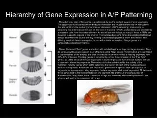
- The adult body plan of Drosophila is established during the earliest stages of embryogenesis. The egg/oocyte itself cannot initiate body plan formation and must therefore rely on instructions that are sent from the mother (remember our discussion of D/V patterning). Instructions for patterning the anterior/posterior axis (in the form of maternal mRNAs and proteins) are provided by a subset of cells from the maternal ovary. As we will see in this lecture many of these mRNAs are localized to specific regions of the embryo. The translated proteins (often transcription factors) will diffuse away from the source thereby forming concentration gradients within the embryo. The differing levels of these transcription factors will activate expression of target genes in a concentration dependent manner. - These “Maternal Effect” genes are tasked with subdividing the embryo into large domains. They do so by activating expression of a set of factors called “Gap” genes. These factors are expressed in large swathes of the embryo and their loss results in an embryo that is missing anywhere from 25%-40% of tissues. The Gap genes in turn directly control the expression of the ”Pair Rule” genes, so called because they are expressed in seven stripes and their removal leads to the loss of tissues in alternating segments. The embryo is further subdivided by the activity of the “Segment Polarity” genes which act to determine the identity of each of the fourteen different embryonic segments. And finally, the “Homeotic” genes confer specific fates upon groups of segments (ie leg segments, wing segments, haltere segments, abdominal segments). Mutations in these genes lead to the transformation of one segment into another. For example, loss of Antennepedia (Antp) leads to the conversion of legs into antennae while overexpression in the antenna will lead to its conversion into a leg. Hierarchy of Gene Expression in A/P Patterning
- In Drosophila the female ovaries (right panel) are located within the abdomen of the adult fly. Each ovary contains a set of sixteen ovarioles. The most anterior tip of the ovariole contains the germarium, the location of the germline stem cells. These stem cells divide and produce cells which will ultimately give rise to the developing egg/oocyte (after fertilization it is called the embryo). There are fourteen distinct stages to the development of each egg/oocyte. At the end of stage 14 the egg/oocyte will first pass through the lateral oviduct, then through the common oviduct and finally through the uterus and vulva. - While the egg/oocyte is developing within the ovariole, it is also being patterned in the dorsal-ventral and anterior-posterior axes. We have already discussed D/V patterning at length. In the lecture we will talk about the initiation of A/P patterning. In contrast to D/V patterning, A/P patterning does not involve the secretion of a ligand. Instead gradients of mRNA and protein localization will be established in the early embryo. This will ultimately lead to differential gene expression along the anterior-posterior axis. Structure of the Drosophila Ovary
- During development of the egg/oocyte a set of ovarian follicle cells (green) will ensheath the developing egg. Remember that is a subset of these cells that secrete the Spatzle ligand during dorsal-ventral patterning. - One cell will give rise to the developing oocyte (the Toll receptor is located within the membrane of the oocyte). - A set of fifteen cells (called nurse cells) will lie adjacent to the anterior quadrant of the oocyte. These cells are connected to each other by structures called ring canals. This allows proteins and mRNA transcripts to be passed between the nurse cells. - The nurse cells are also connected to the oocyte (also by ring canals). During development the nurse cells will deposit large quantities of mRNA transcripts and proteins into the developing egg/oocyte. These are necessary to get development of the embryo started prior to the onset of zygotic transcription. Many of the factors that are deposited from the nurse cells are required for the proper patterning of the anterior-posterior axis of the embryo. - mRNAs that are made in the nurse cells (mother) and deposited in the egg/oocyte (child) are said to be transcribed from “maternal effect” genes. This term is appropriate since transcription of genes in the parent has an effect on the next generation. Please note that the deposition of mRNAs and proteins is not unique to Drosophila – this takes place in all vertebrates including mammals (humans included). Structure of the Drosophila Ovariole
- A large complement of mRNAs and proteins are deposited into the developing oocyte. These factors are manufactured in the adjacent nurse cells (nc) and are dumped into the oocyte through actin-rich ring canals (rc). Three key mRNA transcripts correspond to the bicoid, oskar and gurken genes. These mRNAs are deposited into the anterior pole of the oocyte. One would expect that simple diffusion of these transcripts through the cytoplasm of the oocyte would result in the formation of a concentration gradient in which the highest levels of each transcript would be found at the anterior pole and the lowest levels would be seen at the posterior pole. However, that does not happen. - Instead, all bicoid mRNA transcripts are localized to the anterior pole (top left and below) while the pool of oskar mRNA transcripts is localized to the posterior pole (middle left and below). The gurken mRNA transcript is localized to the anterior dorsal pole (bottom left and below). This localization is important for patterning the embryo in the anterior-posterior axis. In the next slide we will discuss the mechanism by which this localization is achieved. Localization of Maternally Deposited mRNAs maternal bicoid mRNA maternal oskar mRNA maternal gurken mRNA
- The localization of the bicoid, oskar and gurken mRNA transcripts to discrete positions within the developing oocyte is dependent upon the activity of several motor proteins and a microtubule meshwork. - Alpha and beta tubulin subunits are organized into polymers called microtubules. Each microtubule has two polarized ends – a plus end and a minus end. Generally the microtubules within the developing oocyte have their plus ends oriented towards the posterior of the oocyte and their minus ends oriented towards the anterior pole. - Cargo (such as vesicles, proteins and mRNA transcripts) can be transported along the microtubules in a direction dependent manner. This directed movement is dependent upon the motor proteins dynein and kinesin. - Dynein will move cargo towards the minus end of microtubules while kinesin will move cargo towards the plus end of microtubules. From the localization of the mRNA transcripts you can tell that the oskar mRNA transcripts are transported by kinesin to the posterior pole while both bicoid and gurken mRNAs are transported to the anterior pole by dynein. Microtubules and mRNA Localization
- Kinesin is a plus end motor protein therefore it walks towards the plus end of microtubules. It is comprised of several different subunits. One subunit contains an ATPase domain which allows it to generate energy for movement via the hydrolysis of ATP. Kinesins are known to play roles in mitosis, meiosis and in the transport of cargo such as membrane vesicles, proteins and mRNA transcripts such as oskar. In eukaryotes there are over 14 distinct types of kinesins. The division of kinesin proteins into separate classes is based on overall structure and known biological functions. The picture on the right is an artists rendition of a kinesin protein walking a vesicle down a microtubule track. - Dynein is a minus end motor protein thus it walks along microtubules towards the minus end. Like kinesin it is comprised of several different subunits of which one also contains an ATPase domain which is used for energy generation. Dyneins are divided into two classes – cytoplasmic and axonemal. The cytoplasmic dyneins are used to properly position organelles such as the nucleus and Golgi apparatus within the cell. They are also used to transport vesicles, proteins and mRNA transcripts. In contrast the axonemal dyneins are found within cilia and flagella – these are used for the movement of both structures. The picture on the left is an artists rendition of dynein walking down a microtubule towards the minus end. Microtubule Motors – Dynein and Kinesin
- The 3` UTR of bicoid contains a regulatory element that is bound by members of the dynein complex. Since dynein walks cargos to the minus end of microtubules the bicoid mRNA transcript will be transported and localized to the anterior pole of the oocyte (this is where the minus ends are located. Similarly, the 3` UTR of oskar contains a regulatory element that is bound by members of the kinesin complex. As kinesin walks cargo towards the plus end of microtubules, the oskar transcripts will be localized to the posterior pole. If you generate an oskar mRNA that contains the bicoid 3`UTR and inject it into an otherwise normal embryo you will create an embryo that has two tail ends. That is because the endogenous oskar mRNA will be localized properly to the posterior pole while the mutant oskar transcript (that you created) will now localize to the anterior pole and transform that tissue into posterior tissue. - oskar transcripts are not the only ones that are localized to the posterior pole after being deposited into the anterior pole of the oocyte by the nurse cells. nanos mRNA transcripts behave in the same manner. Like oskar transcripts, nanos mRNAs also contain a regulatory element within their 3` UTR that is bound by kinesin motor proteins. If you generate a mutant nanos mRNA that contains the bicoid 3`UTR and inject it into an otherwise normal embryo you will also create an embryo with two tails (see image to the left – right column). - The experiments with oskar and nanos indicate that the correct localization of these transcripts are critical for anterior-posterior patterning. In the next slide we will see why bicoid mRNA must be localized to the anterior pole and why both oskar and nanos transcripts must be localized to the posterior pole. Interaction Between Motor Proteins and mRNAs
- During the early development of the embryo the maternal bicoid and nanos mRNA transcripts will be translated. The Bicoid and Nanos proteins will then diffuse creating classic concentration gradients. The highest levels of Bicoid will be found at the anterior pole while the highest levels of Nanos will be found at the posterior pole. Decreasing amounts of the proteins will be found towards the center of the embryo (see images to the left). - Bicoid is a transcription factor whose roles in development is to activate the expression of two genes: orthodenticle (otd) and hunchback (hb). otd expression is restricted to the regions of the embryo that have the highest levels of Bicoid protein. This is due to the presence of low affinity Bicoid sites within the embryonic enhancer (note the similarities with the twist enhancer). hb expression expands into the middle of the embryo where there are intermediate levels of Bicoid protein. This is due to the presence of both high and low affinity Bicoid sites within the embryonic enhancer (note the similarity with the rho enhancer). Converting Bcd mRNA Transcript Localization into a Protein Gradient
- During early development mRNAs and proteins must be able to move freely through the egg/oocyte. Remember that maternal mRNAs are being localized to the anterior and posterior poles. This requires that microtubule tracks be able to run across the entire embryo unhindered. Also, note that after the maternal mRNA transcripts are translated, the encoded proteins will diffuse away from the mRNA transcript source. In order for this events to happen correctly the early embryo cannot be cellularized. In fact, during the earliest stages of development the embryo is characterized as being a single cell with multiple nuclei (left panel). - As proteins diffuse across the embryo the nuclei are bathed in different concentrations of each factor (ie Bicoid, Oksar and Nanos). These different concentrations must be then maintained so that transcription of the zygotic Gap genes can be activated differentially across the embryo. Just prior to Gap gene transcription the embryo will undergo cellularization (middle and right panels). Cellularization is the process by which the single cell embryo becomes a multicellular organism. This traps the maternal proteins with each cell at a concentration that is appropriate for that cellular position along the A/P axis. For example cells at the most anterior pole will now contain the highest amounts of Bicoid while those at the midsection will have lower levels and cells at the posterior pole will completely lack Bicoid. These differences will lead to the transcription of orthodenticle in the anterior pole and Kruppel in the midsection. Both genes will remain off in the posterior pole. - Note that cellularization of the embryo prevents proteins such as Bicoid, Nanos and Oskar from diffusing any further across the embryo. If cellularization is delayed or prevented then these proteins will continue to diffuse and ultimately their concentration across the embryo will be equal in all cells – the slope of the gradient will be a flat line. Regulating Diffusion Gradients in the Embryo
Within the anterior half of the embryo there are high levels of Bicoid (Bcd - activator) and Hunchback (Hb - genes that are necessary for the formation of the head segments. One gene that is shut off in the anterior section is Kruppel (Kr) – it is required for the formation of the midsection. Even though the Kr embryonic enhancer contains both Bcd and Hb binding sites the activity of the repressor dominates at the higher concentrations. In a bcd mutant Kr expression is activated in the anterior section of the embryo. This in due to the loss of the Hb repressor. It also indicates that other activators (in addition to Bcd) are also used to initiate Kr expression. At the midsection of the embryo the levels of both proteins begin to taper off. Kr expression is activated since Bcd activity dominates over Hb at lower levels. Thus the anterior border of Kr expression is set by the combined activities of Bcd and Hb. The posterior boundary of Kr expression is set by two transcriptional repressors called Knirps (Kni) and Giant (Gt). The expression of these two genes is activated by the gradients that are initiated by oskar and nanos at the posterior pole. The combined efforts of Bcd, Hb, Kni and Gt restricts Kr expression to the embryonic midsection. The Kni and Gt proteins also play important roles in regulating the fate of cells within the posterior section of the embryo. - Hunchback, Kruppel, Knirps and Giant are considered Gap genes since they are expressed in large domains and determine the fates of cells within these domains. Gap Genes Subdivide the Early Embryo Hb KrKni Gt
- Which element within the maternal effect mRNAs allows for localization of the transcripts to discrete positions within the egg/oocyte? - What would happen if a mutation within the bicoid gene disrupted the dynein interaction sequence within the mRNA transcript? - What would happen if a mutation within the bicoid gene changed the affinity of the mRNA transcript from Dynein to Kinesin? - Which feature of the developing egg/oocyte allows for proteins to diffuse and activate gene expression in a concentration dependent manner? - How are microtubules oriented within the developing egg/oocyte? - What would happen if microtubules were depolymerized within the egg/oocyte? - In what directions do Dynein and Kinesin move cargo? - What activity allows for Dynein and Kinesin to walk along microtubules? - What accounts for the different expression patterns of orthodenticle and hunchback? - What happens to zygotic hunchback and Kruppel expression in the absence of Bicoid? - What would happen if knirps and giant expression were lost within the embryo? Molecular Biology Study Questions
Preview of Upcoming Lecture Topics to be Covered Next Time Homeotic Genes and the Body Plan The Homeodomain Co-linearity of the Hox Cluster Disease associated with Mutations in Hox Genes Textbook Chapter Chapter 21 pp. 762-772 Weekly Article(s) “The Evolution of Color Vision” “Ingenious” “Gene Therapy”