Download
1 / 64
640 likes | 769 Views
This presentation provides an in-depth exploration of the mediastinum's anatomy, dividing it into superior, anterior, middle, and posterior sections. Key features include the aortic arch, major vessels, and the esophagus, along with their functions and histological traits. Additionally, the general circulatory system is examined, covering both systemic and pulmonary circuits, heart development, common defects, and cardiac structure. With emphasis on the importance of the thymus in T-cell production and heart mechanics, this resource is essential for understanding complex physiological interactions.

E N D
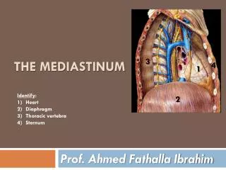
Mediastinum Anatomy & Physiology PA 481 C Tony Serino, Ph.D. Biology Department Misericordia Univ.
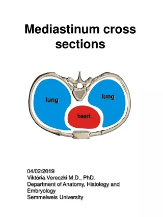
Mediastinum Superior Anterior Middle Posterior Superior and anterior are continuous with each other; both may bereferred to as the superior mediastinum
Superior Mediastinum Transverse thoracic plane Aortic arch Great Vessels of the Heart
Remnant of Ductus arteriosus Ligamentumarteriosum
Usual Aortic Arch Pattern LC RC LS RS BT 65% of all people
Aortic Arch Variations left vert. a. 27% one BT withboth CC exiting 5% 1.2% two BT
SVC Vagus Phrenic BC BC SVC
Structure Order Trachea BC PA Aorta
Esophagus • Function: Deglutition • Two sphincters: upper and lower esophageal sphincters (lower is physiological only) • Retropleural position (therefore, covered by adventitia) • Mucosa: stratified squamous with many mucus glands (esophageal glands) • Muscularis: changes from skeletal to smooth muscle
Thymus Gland • Bilobed organ that is largest in children, but begins to regress sharply at the onset of puberty (around age 11) • It is the site of T-cell lymphocyte production and produces hormones (such as, thymosin) that modifies their physiology
General Circulatory System • Cardiovascular • Consists of a closed system of vessels which transport blood • Two circuits: Systemic and Pulmonary • Arteries move blood away from the heart • Veins move blood toward the heart
General Circulatory System • Lymphvascular –moves lymph • Consist of blind end tubes which collect interstitial fluid (now called lymph) and returns it to circulation • The lymph is cleaned before returned to the blood vessels
Heart as a Dual Pump • Cardiac muscle arranged as whorls that squeeze the blood • Twin pumps: systemic and pulmonary • Four chambers: 2 atria and 2 ventricles
Heart: Anterior View Transverse Pericardial sinus
Heart: Posterior View Oblique Pericardial sinus
Most Common Coronary Arterial Pattern Circumflex a. L. Marginal a. Ant. Desc. a. (LAD) Post. Desc. a. R. Marginal a. Fig. 1.51
Coronary Variation 15% LCA dominant Most people right dominant. (note: which branch gives rise to posterior descending a.determines dominance) Single CA Circumflex from right aortic sinus (4% have an accessory coronary artery)
Coronary Veins Ant. Cardiac veins Great Cardiac v. Coronary sinus Small Cardiac v. Middle Cardiac v. Fig. 1.52
Heart Valves cusps sinus AV (tricuspid) aortic valve (SL) Nodule (corpara aranti)