Download
1 / 152
1.53k likes | 1.65k Views
CHEST AND MEDIASTINUM. CARDIAC. CARDIAC. Coronary Artery Disease Pericardial Effusion Situs Inversus Superior Vena Cava Syndrome. CORONARY ARTERY DISEASE. Description:.

E N D
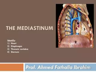
CHEST AND MEDIASTINUM CARDIAC
CARDIAC • Coronary Artery Disease • Pericardial Effusion • Situs Inversus • Superior Vena Cava Syndrome
Description: • Coronary artery disease (CAD) occurs when atherosclerotic plaque (fatty deposits) builds up in the lumen of the coronary arteries. This disease results in a reduced or absent blood flow to the heart.
Etiology: • Atherosclerotic buildup of fatty deposits (plaques) within the lumen of the coronary arteries. This results in a narrowing of the vessel and reduced blood flow.
Epidemiology: • This is the most common type of heart disease affecting men and women.
Signs and Symptoms: • Most common symptom is angina. Other symptoms include shortness of breath, heart palpitation, tachycardia, weakness or dizziness, nausea, and sweating.
Imaging Characteristics: CT • CT angiography (CTA) with IV contrast good to evaluate coronary artery to see if the vessel is occluded. • Calcium scoring without IV contrast shows calcified plaques. MRI • Cardiovascular magnetic resonance (CMR) good to evaluate coronary artery to see if the vessel is occluded.
Treatment: • Interventional procedures (eg. Balloon angioplasty, atherectomy, laser treatment, stent placement) may be used. Coronary bypass surgery may be used in select cases.
Prognosis: • Depends on the patient’s recovery. Change in lifestyle increases better recovery.
Figure 1. Coronary Artery Disease Nonenhanced CT (NECT) shows computer-aided detection and calculation of coronary artery calcification in the right coronary artery (A), left circumflex artery (B), and left anterior descending coronary artery (C).
Figure 2. Coronary Artery Disease Maximum intensity projection CT coronary angiogram showing near-complete occlusion of the right coronary artery.
Description: • This condition is defined as a collection of fluid in the pericardial sac.
Etiology: • Causative factors may include infection, inflammation, immunology, traumatic, uremic, neoplastic, hypothyroidism, chylopericarditis, and congestive heart failure.
Epidemiology: • Majority of cases are related to known or suspected underlying processes which result from secondary diseases.
Signs and Symptoms: • Chest pain, dyspnea, and lightheadedness.
Imaging Characteristics: • Ultrasound is the imaging modality of choice. CT • Good to differentiate between fluid and blood (hemopericardium). • Can detect pericardial calcification. • Normal pericardial thickness is 3 mm. • Pericardial thickness greater than 4 mm is suggestive of pericardial thickening. • Pericarditis may be hyperdense on IV contrast examination.
MRI • Normal pericardial thickness is 3 mm. • Pericardial thickness greater than 4 mm is suggestive of pericardial thickening. • CMR can assess ventricular filling patterns.
Treatment: • Many patients require pericardiocentesis to treat or prevent cardiac tamponade while other patients will need surgical intervention.
Prognosis: • This is a serious condition and patients should be hospitalized until the treatment is accomplished or symptoms improve.
Figure 1. Pericardial Effusion Figure 2. Pericardial Effusion Contrast-enhanced CT (CECT) shows increased fluid density around the heart consistent with a pericardial effusion. Normal for comparison.
Description: • Situs inversus or dextrocardia (cardiac apex pointing to the right) occurs when the morphologic right atrium is located on the left side of the patient, and the morphologic left atrium is located on the right side of the patient. Situs inversus totalis is a complete right-to-left reversal (transposition) of the thoracic and abdominal organs.
Etiology: • Congenital.
Epidemiology: • Occurs in about 0.1% of the population. Situs inversus with dextrocardia is more common and 3% to 5% have sided aortic heart disease. Most of these patients will have a right sided aortic arch. Situs inversus with levocardia is rare and is usually associated with congenital heart disease.
Signs and Symptoms: • Usually asymptomatic. However, some patients, approximately 25%, may have underlying condition called primary ciliary dyskinesia (PCD), also known as Kartagener syndrome. Kartagener syndrome is characterized as situs inversus, bronchiectasis and, chronic sinus infections.
Imaging Characteristics: CT • Preferred modality to diagnosis situsinversus with dextrocardia. • Shows good anatomic detail of organ position and great vessel. MRI • Useful for difficult cases and with associated cardiac anomalies.
Treatment: • No treatment.
Prognosis: • Good.
Figure 1. Situs Inversus CT axial (A) and coronal multiplanar reconstruction (MPR) (B) shows the heart apex on the right.
Description: • This syndrome is the result of an obstruction of blood flow through the superior vena cava (SCV).
Etiology: • May be caused from radiation, cannulation (eg, central venous catheters), tumor bulk, adenopathy, or fibrosing mediastinitis.
Epidemiology: • Majority of cases involve malignant mediastinal tumors.
Signs and Symptoms: • Dyspnea is the most common symptom. Other symptoms include, cough, head and neck fullness, and headache.
Imaging Characteristics: CT • Initial test of choice to determine the cause of an obstruction. • Obstruction of SVC with multiple collateral veins. • SVC thrombosis. • Mediastinal tumor obstructing the SVC. MRI • Good alternative for patients who may be allergic to iodinated contrast or have renal failure.
Treatment: • Depends on the cause. Initial treatment focus is to relieve symptoms. Radiation therapy and chemotherapy may be used to treat tumors. Thrombolytics may be used to treat a thrombus. Surgery may be useful in select cases.
Prognosis: • Primarily depends on the cause and course of treatment.
Figure 1. Superior Vena Cava Syndrome CECT coronal (A) and MPR CECT sagittal (B) images show significant narrowing of the superior vena cava with development of multiple collaterals.
Figure 2. Superior Vena Cava Syndrome Axial CECT shows significant narrowing of the superior vena cava with development of multiple collaterals.
CHEST AND MEDIASTINUM INFECTION
Description: • A fungal infection affecting the pulmonary system.
Etiology: • The Histoplasma capsulatum (H capsulatum) is the fungal organism which causes histoplasmosis. The organism enters the body through the lungs.
Epidemiology: • It is endemic in the Ohio, Missouri, and Missisippi river valley areas. This is the most common endemic fungal infection seen in humans.
Signs and Symptoms: • Majority of patients are asymptomatic. For cases where symptoms occur, they may include: fever, shortness of breath, pneumonia, muscle aches, headaches, chills, and loss of appetite.
Imaging Characteristics: CT • Good to see calcified granulomas in the lung. • Good to see hilar and mediastinallymphadenopathy. • Useful in evaluating patients with mediastinal fibrosis as an ill-defined soft tissue mass surrounding the trachea. • Noncalcifiedgranuloma may be difficult to differentiate from neoplastic lesion.
Treatment: • Antifungal medication may be used.
Prognosis: • Depends on the severity of the disease. As severity of the disease increases, so does the chance that lifelong problems will increase.