Overview of the Mediastinum: Anatomy, Subdivisions, and Clinical Significance
260 likes | 484 Views
The mediastinum is a crucial anatomical region situated between the two pleural sacs, bordered by the sternum in front and the thoracic vertebrae behind. It is divided into superior and inferior compartments, with the inferior subdivided into anterior, middle, and posterior mediastina. Each subdivision contains vital structures, including the heart, major vessels, nerves, and lymph nodes. Understanding the mediastinum's anatomy is essential for diagnosing and managing conditions like mediastinitis and mediastinal tumors.

Overview of the Mediastinum: Anatomy, Subdivisions, and Clinical Significance
E N D
Presentation Transcript
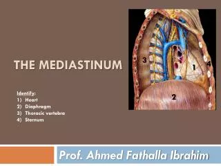
MEDIASTINUM Subdivisions and contents
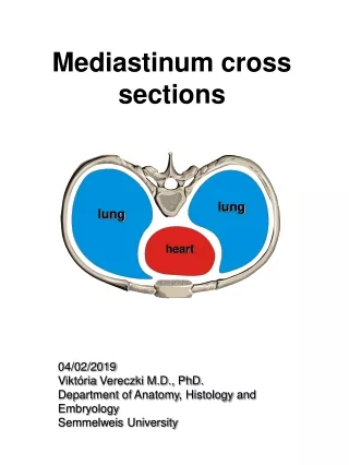
Mediastinum is a central partition between the 2 pleural sacs limited on either sides by the mediastinal pleura • Extent In front – Sternum Behind – Bodies of 12 thoracic vertebrae Above – Thoracic inlet Below – The diaphragm On each side – Mediastinal pleura
Subdivisions An imaginary horizontal plane extending from the sternal angle to the lower border of T4 into superior and inferior
Inferior mediastinum is further divided by the pericardial sac into anterior, middle and posterior
Superior mediastinum • Boundaries In front – Manubrium sterni Behind – Bodies of upper 4 thoracic vertebrae, intervertebral discs and anterior longitudinal ligament Above – Thoracic inlet Below – The diaphragm On each side – Mediastinal pleura
Anterior mediastinum • Boundaries In front – Body of the sternum Behind – Pericardium Above – Imaginary horizontal plane extending from sternal angle to the lower border of T4 Below – The diaphragm On each side – Mediastinal pleura
Contents • Superior and inferior sternopericardial ligaments • Loose areolar tissue • Retrosternal lymph nodes • Mediastinal branches of internal thoracic artery
Middle mediastinum • Widest subdivision • Occupied mainly by the heart and pericardium • Limited on each side by mediastinal pleura
Contents • Heart enclosed in the pericardium • Arteries – Ascending aorta and pulmonary trunk dividing into right and left pulmonary arteries • Veins – Lower part of superior venacava, arch of azygos vein and 4 pulmonary veins • Nerves – phrenic, deep cardiac plexus • Lymph nodes – inferior tracheobronchial nodes • Tubes – bifurcation of trachea, right and left bronchi
Contents of middle mediastinum Ascending aorta Phrenic nerve Deep cardiac plexus Left pulmonary artery Left bronchus Pulmonary veins Heart within pericardium
Posterior mediastinum • Boundaries In front (from above downwards) – Bifurcation of trachea, pulmonary vessels, fibrous pericardium, posterior sloping surface of the diaphragm Behind – Bodies of lower 8 thoracic vertebrae, intervertebral discs and anterior longitudinal ligament Above – Imaginary horizontal plane Below – The diaphragm On each side – Mediastinal pleura
Thoracic duct Left recurrent laryngeal nerve • Contents Aorta and its branches Left vagus Azygos vein oesophagus
Oesophagus Arteries – Descending thoracic aorta and its branches Veins – Azygos, Hemiazygos, Accessory hemiazygos Nerves – Vagus, splanchnic nerves Lymph nodes – posterior mediastinal nodes Thoracic duct
Surgical importance / Applied anatomy • Mediastinitis – Structures of mediastinum are embedded in loose connective tissue. The posterior mediastinum is continuous with the neck through the superior mediastinum. Therefore infection from these regions may spread to superior and posterior mediastina • Deflection of mediastinum – In pneumothorax, the lung on that side immediately collapses and mediastinum is deflected to opposite side the patient suffers from breathlessness & upon examination trachea and heart displaced to opp side
All large veins of sup medaistinum are located on the right side. Therefore during increased venous return, the veins expand whereas the arteries do not expand at all. Thus there is much dead space on the right, and hence tumours or fluids tend to project on this side. • Mediastinal tumours • Mediastinal syndrome – compression of mediastinal structures by any growth or tumour
Mediastinoscopy – surgical procedure to look at the organs, tissues and lymph nodes between the lungs for abnormal areas. An incision is made at the top of the breastbone and endoscope is inserted into the chest. Tissue and lymph nodes may be taken by biopsy.