Mediastinum
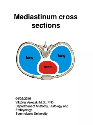
Mediastinum . Mediastinum . central structures between two pleural sacs, sternum & vertebral bodies . Mediastinum. Region 1. superior mediastinum: behind manubrium (pleura are apart here) thymus gland - most anterior structure, behind manubrium in front of brachiocephalic vein, aortic arch


Mediastinum
E N D
Presentation Transcript
Mediastinum • central structures between two pleural sacs, sternum & vertebral bodies
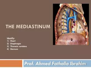
Mediastinum • Region • 1. superior mediastinum: behind manubrium (pleura are apart here) • thymus gland - most anterior structure, behind manubrium • in front of brachiocephalic vein, aortic arch • slowly shrinks w age (largest in childhood), replaced largely by adipose • 2. inferior: separation from superior is at line between sternal angle & intervertebral disc 4- 5a. anterior - in front of pericardium (thin space) b. middle - contains pericardium, phrenic nerves, blood vesselsc. posterior - behind pericardium
Vessels • arch of the aorta - opposite arch of azygos, just superior to root of left lung • thoracic aorta: ascending - arch - descending aorta - posterior to root of right lung, arteries exit superiorly from arch: • brachiocephalic artery • brachiocephalic artery then divides to right common carotid and right subclavian aretry • Left common carotid • Left subclavian artery
1. Brachiocephalic Artery 2. Carotid Artery, Left 3. Subclavian Artery, Left 4. Aortic Arch 5. Ascending Aorta 6. Pulmonary Trunk 7. Pulmonary Artery, Left 8. Pulmonary Vein, Left 9. Atrim, Left 10. Bicuspid Valve 11. Aortic Semilunar Valve 12. Ventricle, Left 13. Interventricular Septum 14. Ventricle, Right 15. Papillary Muscle 16. Chordae Tendinae 17. Tricuspid Valve 18. Atrium, Right 19. Pulmonary Semilunar Valve 20. Superior Vena Cava
Veins • superior & inferior vena cava - entering the right atrium = junction of brachiocephalics • brachiocephalic veins - continuous with superior vana cava superiorly, behind sternoclavicular joint • a. combination of subclavian & internal jugular veins • b. internal thoracic veins also drain into brachiocephalics
Blood vessels • Pulmonary trunk • Pulmonary trunk: short, just left of aortic arch - branches to Right, Left pulmonary artery- ligamentum arteriosum - residual of ductus arteriosum - connects to aorta in fetal circulation • Azygos vein
Other structures • Other structures in mediastinum • Trachea - behind right brachiocephalic vein • Esophagus - behind trachea
Nerves • phrenic nerve: in mediastinal pleura on each side, anterior to roots of lungs • anterior to subclavian artery, parallels internal thoracic artery • adjacent to brachiocephalic veins, superior vana cava or aorta, pericardium; to diaphragm • vagus nerves: posterior to phrenics • Right vagus - between brachiocephalic artery & vein • Left vagus - between left common carotid & subclavian artery • recurrent laryngeal branch of left vagus - curls under aortic arch behind ligament arteriosum • recurrent laryngeal branch of right vagus - curls under the subclavian artery
Plexus from Vagus • Cardiac plexus: 2 portions • superficial - just under aortic arch; & deep - posterior to arch, anterior to trachea (gets most branches) comprised of branches of sympathetic trunks: • 3 cervical cardiac branches - from neck, thru inlet to superior Mediastinum • several thoracic branches • Pulmonary plexuses: 4 - R & L anterior & posterior • in front & behind pulmonary artery on each side, at root of lungs • from there into lungs • nerve filaments arise as branches from cardiac plexus • Coronary plexus - nerve filaments also come from cardiac plexus - to heart, follow coronary arteries
Pericardial Sac • Serous Pericardium - serous membrane - double layer • a. visceral pericardium - forms epicardium - outer covering of heart • b. parietal pericardium - thicker, tougher than pleura • c. pericardial cavity • Fibrous pericardium - dense fibrous CT - fused with parietal serous pericardium continuous with deep fascia of neck also attaches to diaphragm below & to esophagus & aorta posteriorly