MEDIASTINUM
490 likes | 662 Views
The mediastinum is a crucial partition between the right and left pleural sacs, encompassing all structures within the thoracic cavity's intermediate compartments. It can be divided into superior and inferior mediastinum. The superior mediastinum contains the esophagus, trachea, arch of the aorta, brachiocephalic veins, and thymus gland. The inferior mediastinum is further categorized into anterior, middle, and posterior sections, housing vital components like the heart, pericardium, and vessels. Each section is bounded by specific anatomical landmarks and contains essential nervous and lymphatic structures.

MEDIASTINUM
E N D
Presentation Transcript
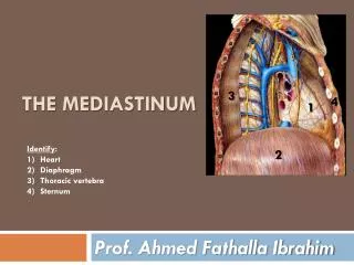
MEDIASTINUM Dr. Ahmed Fathalla Ibrahim
DEFINITION OF MEDIASTINUM • It is a partition between the right & left pleural sacs. It includes all the structures which lie in the intermediate compartments of the thoracic cavity
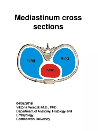
DIVISIONS OF MEDIASTINUM • It is divided by a horizontal plane extending from sternal angle to lower border of 4th thoracic vertebra into: • Superior mediastinum: above the plane • Inferior mediastinum: below the plane, it is subdivided into: • Anterior mediastinum: in front of pericardium • Middle mediastinum: contains heart & pericardium • Posterior mediastinum: behind pericardium
SUPERIOR MEDIASTINUM • BOUNDARIES: • Anterior: manubrium sterni • Posterior: Upper 4 thoracic vertebrae • Superior: Plane of thoracic inlet • Inferior: Horizontal plane • On each side: Pleura
SUPERIOR MEDIASTINUM • CONTENTS: • FROM BEHIND FORWARD: • Esophagus • Trachea • Arch of aorta & its 3 branches: brachiocephalic, left common carotid & left subclavian arteries • Right & left brachiocephalic veins & superior vena cava • Thymus gland
SUPERIOR MEDIASTINUM • OTHER CONTENTS: • Nerves: • Right & left vagus • Right & left phrenic • Right & left sympathetic trunks • Left recurrent laryngeal • Lymphatic structures: • Thoracic duct • Lymph nodes
POSTERIOR MEDIASTINUM • BOUNDARIES: • Anterior: Pericardium & diaphragm • Posterior: Lower 8 thoracic vertebrae • Superior: Horizontal plane • Inferior: Diaphragm • On each side: Pleura
POSTERIOR MEDIASTINUM • CONTENTS: • Esophagus (most anterior structure) • Thoracic duct • Right & left vagus • Descending aorta • Azygos & hemiazygos veins • Right & left sympathetic trunks & their branches (splanchnic nerves) • Lymph nodes
MIDDLE MEDIASTINUM • CONTENTS: • Pericardium & heart • Arteries: ascending aorta, pulmonary trunk • Veins: lower half of superior vena cava, terminations of inferior vena cava & pulmonary veins • Nerves: phrenic • Lymph nodes
VEINS • BRACHIOCEPHALIC:(Superior mediastinum) • FORMATION: by union of internal jugular & subclavian vein (behind medial end of clavicle) • END: Both veins unite to form S.V.C. • RIGHT VEIN:shorter & has a vertical course, related laterally to right phrenic nerve & right pleura & lung, itstributaries in thorax: right 1st posterior intercostal vein, right internal thoracic vein, right lymphatic duct • LEFT VEIN:longer & has an oblique course, relatedanteriorly to manubrium & thymus gland, & posteriorly to branches of arch of aorta, its tributaries in thorax: left 1st posterior intercostal vein, left superior intercostal vein, left internal thoracic vein, thoracic duct
VEINS • SUPERIOR VENA CAVA:(Superior & middle mediastinum) • FORMATION: by union of brachiocephalic veins, behind lower border of right 1st costal cartilage • END: opens into right atrium behind right 3rd costal cartilage • TRIBUTARIES: azygos vein
VEINS • AZYGOS VEIN:(Posterior mediastinum) • ORIGIN: by union of right ascending lumbar & subcostal veins (passes through aortic opening of diaphragm) • END: forms an arch above the root of right lung & ends in S.V.C. opposite lower border of T4 • RELATIONS: • Anterior: esophagus • Posterior: thoracic vertebra • Right: right pleura & lung • Left: thoracic duct • TRIBUTARIES: superior & inferior hemiazygos veins, right superior intercostal vein, right posterior intercostal veins (from 4th to 11th), right bronchial veins, esophageal & pericardial veins
VEINS • INFERIOR HEMIAZYGOS:(Posterior mediastinum) • ORIGIN: by union of left ascending lumbar & subcostal veins (passes through left crus of diaphragm) • END: into azygos vein, opposite T8 • TRIBUTARIES: left posterior intercostal veins (9th to 11th), esophageal veins
VEINS • SUPERIOR HEMIAZYGOS:(Posterior mediastinum) • ORIGIN: by left posterior intercostal veins (4th to 8th) • END: into azygos vein, opposite T7 • TRIBUTARIES: left bronchial veins INFERIOR VENA CAVA:(Posterior mediastinum) • END: passes through vena caval opening of diaphragm &opens into right atrium behind right 6th costal cartilage
ARTERIES • AORTA: • ASCENDING AORTA: (Middle mediastinum) • ORIGIN: at the base of left ventricle opposite lower border of left 3rd costal cartilage • END: ascends upward, forward & to the right & continues as arch of aorta • BRANCHES: right & left coronary arteries
ARTERIES • ARCH OF AORTA:(Superior mediastinum) • ORIGIN: continuation of ascending aorta, opposite upper border of right 2nd costal cartilage • COURSE & RELATIONS: ascends upward backward & to the left(behind manubrium & in front of trachea) then curves backward (to the left of trachea) then finally curves downward • TERMINATION: continues as descending aorta, opposite lower border of T4
ARTERIES • BRANCHES OF ARCH OF AORTA:(Superior mediastinum) • BRACHIOCEPHALIC: ascends upward & to the right(behind left brachiocephalic vein & in front of trachea) & divides into right common carotid & right subclavian arteries (behind right sternoclavicular joint) • LEFT COMMON CAROTID: ascends upward & to the left(to the left side of brachiocephalic artery) & enters the neck (behind left sternoclavicular joint) • LEFT SUBCLAVIAN: ascends upward (behind left common carotid artery, in front of esophagus, to the left side of trachea), arches over apex of left lung to enter neck
ARTERIES • DESCENDING AORTA:(Posterior mediastinum) • ORIGIN: continuation of arch of aorta • TERMINATION: passes through aortic opening of diaphragm (opposite T12) & continues as abdominal aorta • RELATIONS: • Anterior: esophagus • Posterior: thoracic vertebrae • Right: thoracic duct • Left: left pleura & lung • BRANCHES: posterior intercostal (from 3rd to 11th), subcostal, bronchial, esophageal, pericardial arteries
ARTERIES • PULMONARY TRUNK (Middle mediastinum) • ORIGIN: from upper part of right ventricle, behind sternal end of left 3rd costal cartilage • COURSE: ascends upward & to the left & divides (at lower border of T4) into: • Right pulmonary:runs behind ascending aorta & S.V.C to enter root of right lung • Left pulmonary:runs in front of desending aorta to enter root of leftlung
TRACHEA • BEGINNING: continuation of larynx, opposite C6 • TERMINATION: bifurcates into 2 bronchi, opposite lower border of T4 • RELATIONS: (in superior mediastinum) • Anterior: arch of aorta, brachiocephalic & left common carotid arteries • Posterior: left recurrent laryngeal nerve, esophagus • Right: right vagus nerve • Left: arch of aorta, left subclavian artery • NERVE SUPPLY: sympathetic trunks & vagus • BLOOD SUPPLY: inferior thyroid vessels • LYMPHATIC DRAINAGE: pretracheal & paratracheal
ESOPHAGUS • BEGINNING: continuation of pharynx, opposite C6 • TERMINATION: passes through esophageal opening of diaphragm (opposite T10) & joins stomach • RELATIONS: (in superior mediastinum) • Anterior: left recurrent laryngeal nerve, trachea, left subclavian artery • Posterior: thoracic vertebrae • Right: right pleura & lung • Left: thoracic duct, left pleura & lung
ESOPHAGUS • RELATIONS: (in posterior mediastinum) • Anterior: pericardium, separating it from left atrium • Posterior: thoracic duct, descending aorta, azygos vein • Right: right pleura & lung • Left: descending aorta, left pleura & lung • NERVE SUPPLY: as trachea • ARTERIAL SUPPLY: descending aorta • VENOUSDRAINAGE: azygos & hemiazygos • LYMPHATIC DRAINAGE: posterior mediastinal lymph nodes
THORACIC DUCT • ORIGIN: from upper end of cysterna chyli (opposite L1 & L2) • COURSE: passes through aortic opening of diaphragm, ascends in posterior mediastinum (behind esophagus) & in superior mediastinum (to the left of esophagus) to enter root of neck • END: in left brachiocephalic vein • RELATIONS: ( in posterior mediastinum) • Anterior: esophagus • Posterior: thoracic vertebrae • Right: azygos vein • Left: descending aorta
THORACIC DUCT • TRIBUTARIES: • It drains lymph from both sides of the body below the diaphragm through cysterna chyli • It drains lymph from left half of the body above diaphragm through: • Left jugular lymph trunk: drains left side of head & neck • Left subclavian lymph trunk: drains left upper limb • Left bronchomediastinal lymph trunk: drains left side of thorax
RIGHT LYMPHATIC DUCT • ORIGIN:formed by union of: • Right jugular lymph trunk: drains right side of head & neck • Right subclavian lymph trunk: drains right upper limb • Right bronchomediastinal lymph trunk: drains right side of thorax • END:in right brachiocephalic vein
NERVES • PHRENIC NERVES:(Superior & middle mediastinum) • ORIGIN: anterior rami of C3,4,5 • COURSE & RELATIONS IN THORAX: • RIGHT: descends to the right side of: right brachiocephalic vein, S.V.C., pericardium, I.V.C. • LEFT: descends to the left side of: arch aorta, pericardium • BRANCHES: • Motor branches to: diaphragm • Sensory branches from: • Mediastinal & central part of diaphragmatic pleura • Fibrous pericardium & parietal layer of serous pericardium • Peritoneum covering central part of undersurface of diaphragm
NERVES • VAGUS NERVES:(Superior & posterior mediastinum) • ORIGIN: 10th cranial nerve • COURSE & RELATIONS IN THORAX: • RIGHT: descends to the right side of: trachea, behind root of right lung (pulmonary plexus), behind esophagus (esophageal plexus), passes through esophageal opening of diaphragm to reach posterior surface of stomach • LEFT: descends to the left side of: arch aorta, behind root of left lung (pulmonary plexus), in front of esophagus (esophageal plexus), passes through esophageal opening of diaphragm to reach anterior surface of stomach
NERVES • BRANCHES IN THORAX: • BOTH VAGI: to lungs & esophagus • RIGHT VAGUS: to heart • LEFT VAGUS: left recurrent laryngeal nerve: curves below arch of aorta, behind ligamentum arteriosum, ascends in groove between trachea & esophagus to reach the neck. It supplies: heart, trachea, esophagus (in thorax) & larynx (in neck)
NERVES • THORACIC PART OF SYMPATHETIC TRUNKS:(Superior & posterior mediastinum) • BEGINNING: the cervical part continues as thoracic part by passing in front of neck of first rib • TERMINATION: the thoracic part continues as lumbar part by passing behind medial arcuate ligament • COURSE: • In upper part of thorax: descend in front of heads of ribs • In lower part of thorax: descend on the sides of bodies of vertebrae • GANGLIA: usually 11 (1st thoracic ganglion fuses with inferior cervical ganglion forming stellate ganglion)
NERVES • BRANCHES: • Rami communicantes: each ganglion receives a white ramus (preganglionic) & gives a grey ramus (postganglionic) to corresponding thoracic spinal nerve • Visceral branches (postganglionic)to thoracic organs (from upper 5 ganglia): to heart, lungs, esophagus, descending aorta • Visceral branches (preganglionic) to abdominal organs: • Greater splanchnic nerve (from 5th to 9th ganglia) • Lesser splanchnic nerve (from 10th 7 11th ganglia) • Lowest splanchnic nerve (from 12th ganglion)