Download
1 / 22
240 likes | 784 Views
Anatomy of the cell and cell division. Exercise #3 Page : #25. Introduction to cells. Cells are very smallest living units in the human body. Cells diameter is only about 0.1 mm. Microscope were invented in the 17 th century.

E N D
Anatomy of the cell and cell division Exercise#3 Page: #25
Introduction to cells • Cells are very smallest living units in the human body. • Cells diameter is only about 0.1 mm. • Microscope were invented in the 17th century. • Robert Hook created the term cell because they reminded him the smalls rooms in a monastery or prison. It was in 1665.
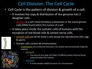
CELLS • The human body contains trillions of cells. • Cytology is the study of cellular structure that incorporated aspects of biology, chemistry and physics. • The human body has 2 general classes of cells. • Sex cells (reproductive cells) • Somatic cells (soma, body) they are all the other cells in the human body.
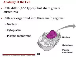
Cell and extracellular fluid • Extracellular fluid is the watery medium that surrounds the cell. • It is also called interstitial fluid. • Interstitial means something standing between. • Cytoplasm includes all the cell content. • The cytoplasm has 2 components: • 1- cytosol (liquid). • 2- organelles(intracellular structures).
Cell membrane: isolation, protection, sensitivity, support, • entrance & exits. • Cytosol: Distributes material by diffusion. • Non membranous organelles • Cilia: movement of materials over the cell surface • Microvilli: increase surface area for absorption • Centrosome: movement of chromosomes during cell division; organization of microtubules and cytoskeleton. • Ribosomes:protein synthesis. • Proteasomes: breakdown and recycling of damaged or abnormal intracellular proteins.
Membranous organelles • Mitochondria: Produce 95% of ATP (energy) • Endoplasmic reticulum • Rough (ER): modification and packaging of proteins. • Smooth (SER): lipids and carbohydrates synthesis. • Golgy apparatus: storage , alteration and packaging of secretory products and lysosomal enzymes. • Lysosomes: intracellular removal of damaged organelles or pathogens.
Membranous organelles • Peroxisome:catabolism of fats; neutralization of toxic compounds • Nucleus: control of protein synthesis, cell metabolism, storage & processing genetic information. • Nucleolus: site for rRNA synthesis & assembly of • ribosomal subunits • Flagellum (only in sperm): movement of the cell.
Golgy Apparatus ALFONSO A. PINO MD.
CELL STRUCTURE AND FUNCTIONS(Martini, pg 65) table 1 • Cell membrane: isolation, protection, sensitivity, support, • entrance & exits • Nucleus: control of protein synthesis, storage & • processing genetic information • Cilia: movement of materials over the cell surface • Mitochondria: produce 95% of ATP (energy) • Rough endoplasmic reticulum: modification & packaging of • new synthesized proteins • Smooth endoplasmic reticulum:lipids & carbohydrate • synthesis • Golgi apparatus:storage, alteration & packaging of • secretory products & lysosomal enzymes
CELL STRUCTURE AND FUNCTIONS(Martini, pg 65) table 1 • Lysosome: Intracellular removal of damaged organelles • & pathogens • Ribosome: protein synthesis • Flagella: cell movement (only in sperm) NOT IN TABLE • Centriole: movement of chromosomes, organization of • microtubules & cytoskeleton • Microvilli: increase surface area for absorption • Peroxisome: neutralization of toxic compounds • Nucleolus: site for rRNA synthesis & assembly of • ribosomal subunits
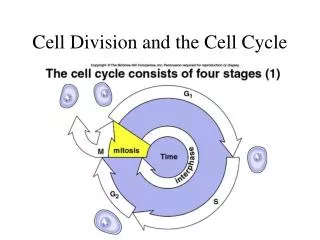
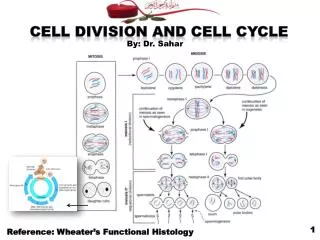
CELL STRUCTURE AND MITOSIS • MITOSIS: • It is the process of nuclear division of the cell • The DNA is separated into two separate but equal sets • Each cell has the same genetic makeup as all its predecessors
CELL STRUCTURE AND MITOSIS(Martini pg 97) • Interphase • 4 stages of mitosis: • Prophase • Metaphase • Anaphase • Telophase
STAGE INTERPHASE Early pr ophase Late pr ophase (prometaphase) Nucleus Spindle fibers Centriole Mitosis begins Centrioles (two pairs) Chromosome with two sister chromatids Astral rays INTERPHASE The chromosomal material cannot be seen Duplication of the DNA occurs During most of the cell cycle the cell is in interphase • The chromosomal material cannot be seen • Duplication of the DNA occurs • During most of the cell cycle the cell is in interphase ALFONSO A. PINO MD.
STAGE INTERPHASE Early pr ophase Late pr ophase (prometaphase) Nucleus Spindle fibers Centriole Mitosis begins Centrioles (two pairs) Chromosome with two sister chromatids Astral rays PROPHASE The 1st stage of Mitosis Nuclear membrane disappear Chromosomes become visible and the mitotic spindle begins to form • The 1st stage of Mitosis • Nuclear membrane disappear • Chromosomes become visible and the mitotic spindle begins to form
STAGE STAGE STAGE CYTOKINESIS Metaphase Anaphase elophase T Chromosomal microtubule Daughter chromosomes Separation Cleavage furrow Daughter cells Metaphase plate METAPHASE • When the spindle fibers are attached to the centromere region of all the chromosomes and chromosomes have been pulled into the same plane of the cell the metaphase stage is reached • When the spindle fibers are attached to the centromere region of all the chromosomes and chromosomes have been pulled into the same plane of the cell the metaphase stage is reached
STAGE STAGE STAGE CYTOKINESIS Metaphase Anaphase elophase T Chromosomal microtubule Daughter chromosomes Separation Cleavage furrow Daughter cells Metaphase plate ANAPHASE • Begins when the 2 daughter stands of each chromosome separate and are pulled towards opposite ends of the cell • Begins when the 2 daughter stands of each chromosome separate and are pulled towards opposite ends of the cell
STAGE STAGE STAGE CYTOKINESIS Metaphase Anaphase elophase T Chromosomal microtubule Daughter chromosomes Separation Cleavage furrow Daughter cells Metaphase plate TELOPHASE It is the last stage of Mitosis The 2 sets of chromosomes are at opposite sides of the cell, and the nuclear membrane has formed • It is the last stage of Mitosis • The 2 sets of chromosomes are at opposite sides of the cell, and the nuclear membrane has formed
STAGE STAGE STAGE CYTOKINESIS Metaphase Anaphase elophase T Chromosomal microtubule Daughter chromosomes Separation Cleavage furrow Daughter cells Metaphase plate CYTOKINESIS • The cell divides into 2 new cells. It happens while the nucleus is in telophase • The cell divides into 2 new cells. It happens while the nucleus is in telophase