Download

1 / 37
380 likes | 658 Views
Chromosomes, the Cell Cycle, and Cell Division. Systems of Cell Reproduction. Four events occur before and during cell division: A signal to reproduce must be received. Replication of DNA and vital cell components must occur. DNA must be distributed to the new cells.

E N D
Systems of Cell Reproduction • Four events occur before and during cell division: • A signal to reproduce must be received. • Replication of DNA and vital cell components must occur. • DNA must be distributed to the new cells. • The cell membrane or cell wall must separate the two new cells.
Systems of Cell Reproduction • Prokaryotes divide by fission. • Most prokaryotes have one circular chromosome. • As DNA replicates, each of the two resulting DNA molecules attaches to the plasma membrane. • As the cell grows, new plasma membrane is added between the attachment points, and the DNA molecules are moved apart. • Cytokinesis separates the one cell into two, each with a complete chromosome.
Systems of Cell Reproduction • Eukaryotic cells divide by mitosis or meiosis. • Three Steps: • The replication of the DNA within the nucleus • The packaging and segregation of the replicated DNA into two new nuclei (nuclear division) • The division of the cytoplasm (cytokinesis)
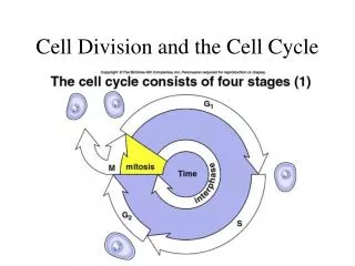
Interphase and the Control of Cell Division • The cell cycle has two phases: mitosis and interphase. • A typical eukaryotic cell will spend most of its life in interphase, the period between divisions of the cytoplasm. • Some cells lose the capacity to divide altogether and stay in interphase indefinitely, while other cells divide regularly or occasionally. A Lily cell in interphase
Interphase and the Control of Cell Division • Interphase consists of three subphases: • G1 (Gap 1): period just after mitosis and before the beginning of DNA synthesis. • S (synthesis): the time when the cell’s DNA is replicated. • G2 (Gap 2): the time after S and prior to mitosis.
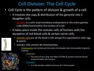
Eukaryotic Chromosomes • Chromatin: the basic unit of the eukaryotic chromosome; a gigantic, linear, double-stranded molecule of DNA complexed with many proteins. • Chromatids: what each chromosome consists of after the DNA of a chromosome replicates during S phase. Centromere
Eukaryotic Chromosomes • Chromosomes are wrapped around proteins called histones. • These wraps of DNA and histone proteins are called nucleosomes. • The core of a nucleosome contains eight histone molecules, two each from four of the histone classes. • During mitosis and meiosis, the chromatin becomes even more coiled and condensed.
Mitosis: Distributing Exact Copies of Genetic Information • Centrosomes: • Replicated when the cell enters S phase and DNA is replicated. • Separate from each other during G2→M. • The orientation determines the cell’s plane of division. • Regions where microtubules form. Parent daughter pairs of centrioles before migration.
Mitosis: Distributing Exact Copies of Genetic Information • Prophase marks the beginning of mitosis. • Chromosomes compact and coil, becoming more dense and visible. • Polar microtubules form between the two centrosomes and make up the developing spindle. • The mitotic spindle serves as a “railroad track” along which chromosomes will move later in mitosis. • Late in prophase, the kinetochores develop in the region around the centromere and are the sites where microtubules attach to the chromatids.
Mitosis: Distributing Exact Copies of Genetic Information • During metaphase, the kinetochores arrive at the equatorial plate. • Chromosomes are fully condensed. • Unraveling of the interconnected DNA molecules at the centromere occurs. Cell in metaphase.
Mitosis: Distributing Exact Copies of Genetic Information • Anaphase begins when the centromeres separate. • Molecular motors at the kinetochores move the chromosomes toward the poles. • Also, the microtubules are shortened at the poles, and the mitotic centers further separate. Cells in mid and late anaphase.
Mitosis: Distributing Exact Copies of Genetic Information • Telophase begins when the chromosomes finish moving. • Nuclear envelopes and nucleoli coalesce and re-form. • Animal cells divide by a furrowing (a “pinching in” or constriction) of the plasma membrane. Cell in late telophase. Sea urchin egg at cytokinesis.
Reproduction: Asexual and Sexual • Mitosis by repeated cell cycles can give rise to vast numbers of genetically identical cells. • Meiosis results in just four progeny, which usually do not further duplicate. The cells can be genetically different.
Reproduction: Asexual and Sexual • Sexual reproduction involves meiosis. • Two parents each contribute a set of chromosomes in a sex cell or gamete. • Gametes fuse to produce a single cell, the zygote. • This creates variety among the offspring beyond that attributed to mutations or the environment.
Reproduction: Asexual and Sexual Homologous Chromosomes • In multicellular organisms, somatic cells each contain two sets of chromosomes. • One comes from each of the two parents. • The members of the pair are called homologous chromosomes and have corresponding but generally not identical genetic information. A a Eye Color b B Hair Color Hairline C c Ear lobe attachment d D
Figure 9.12 Fertilization and Meiosis Alternate in Sexual Reproduction (Part 2) * When haploid gametes fuse in fertilization, they create the diploid zygote (2n). * Haploid cells contain just one homolog of each pair (n).
Reproduction: Asexual and Sexual • Cells in metaphase can be killed and prepared in a way that spreads the chromosomes around a region on a glass slide. • The images of each chromosome can be organized based on size, number, and shape = karyotype.
Meiosis: A Pair of Nuclear Divisions • Meiosis consists of two nuclear divisions that reduce the number of chromosomes to the haploid number. • The DNA is replicated only once. • The functions of meiosis: • To reduce the chromosome number from diploid to haploid. • To ensure each gamete gets a complete set of chromosomes. • To promote genetic diversity among products.
Meiosis: A Pair of Nuclear Divisions • Meiosis I is preceded by an interphase in which DNA is replicated. • During prophase I, synapsis occurs: The two homologs are joined together by a complex of proteins. • This forms a tetrad, or bivalent, which consists of two homologous chromosomes with two sister chromatids.
Meiosis: A Pair of Nuclear Divisions • At a later point, the chromosomes appear to repel each other except at the centromere and at points of attachments which appear x-shaped. • These attachment points reflect the exchange of genetic material between homologous chromosomes, a phenomenon called crossing-over. • This crossing-over increases genetic variation by reshuffling the genes on the homologs.
Figure 9.16 Crossing Over Forms Genetically Diverse Chromosomes
Meiosis: A Pair of Nuclear Divisions • The homologous chromosomes separate in anaphase I. • The individual chromosomes are pulled to the poles, with one homolog of a pair going to one pole and the other homolog going to the opposite pole.
Meiosis: A Pair of Nuclear Divisions • The second meiotic division separates the chromatids. • Meiosis II is similar to mitosis but one difference is that DNA does not replicate before meiosis II. • The number of chromosomes in the resulting cells is therefore half that found in diploid mitotic cells. • In meiosis II, sister chromatids are not identical and there is no crossing-over.
Meiosis: A Pair of Nuclear Divisions • Meiosis leads to genetic diversity. • Synapsis and crossing-over during prophase I mix genetic material of the maternal with that of the paternal homologous chromosomes. • Which member of a homologous pair segregates or goes to which daughter cell at anaphase I is a matter of chance. This phenomenon is called independent assortment. Metaphase I Anaphase I
Meiotic Errors • Nondisjunction occurs when homologous chromosomes fail to separate during anaphase I, or sister chromatids fail to separate during anaphase II. • The result is a condition called aneuploidy.