Cell Division and Cell Cycle
260 likes | 533 Views
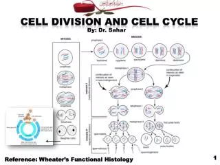
Cell Division and Cell Cycle. By: Dr. Sahar. 1. Reference: Wheater’s Functional Histology. Cell Division. * All multicellular organisms originally develop from a single cell which is the fertilized ovum. * This development includes: Cellular replication. Growth.

Cell Division and Cell Cycle
E N D
Presentation Transcript
Cell Division and Cell Cycle By: Dr. Sahar 1 Reference: Wheater’s Functional Histology
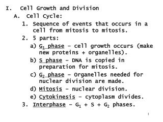
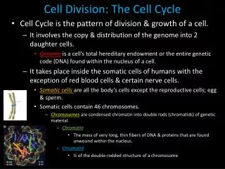
Cell Division *All multicellular organisms originally develop from a single cell which is the fertilized ovum. *This development includes: Cellular replication. Growth. Progressive specialization to do the various body functions. *Cellular replication depends on formation of new cells by the division of the preexisting ones. *There are two kinds of cell division: 1- Mitosis: occurs in all body cells except male and female germ cells. 2- Meiosis: confined to the germ cells that develop into ova and spermatozoa. 2
Mitosis *Definition: Is the process of somatic cell division that results in the formation of two daughter cells genetically identical to each other and to the mother cell. • *Genetically identical= contains the same chromosomal number characteristic to the species. In the human = 46. *Following mitosis, the daughter cells enter a period of growth and metabolic activity prior to further mitotic division. *This period between successive divisions is called the interphase. 4
*Events: Mitotic division includes 2 successive events: 1- Karyokinesis or mitosis= Division of the genetic material (=chromosomes) that has been previously duplicated during the interphase. Chromosomes are then distributed equally between 2 potential daughter cells. 2- This is followed by division of the cytoplasm (cytokinesis). Chromosomes during mitosis 5 Events of mitotic division
*Phases: Mitosis is a continuous process, but, for descriptive purpose, its events are described into 4 phases: 1- Prophase. 2- Metaphase. 3- Anaphase 4- Telophase. 6
The two round objects above the nucleus are the centrosomes. Thick , coiled chromosomes, each with two chromatids, are lined up on the metaphase plate (At the equatorial plane). Note the decondensing chromosomes. 7
Meiosis *Definition: Is a process of division in the germ cell in which the number of chromosomes is reduced from the diploid number, typical of somatic cells (46), to a haploid number in the gametes (23 in sperms and ova). *The diploid number is then restored by fusion of the sperm and ovum at fertilization. *Reduction of chromosome number is accomplished by occurrence of two cell divisions of which only the first is preceded by duplication (= replication) of chromosomes. 10
*Events: A diploid cell contains a full set of chromosome pairs, each pair containing one chromosome from each parent. These chromosome pairs are called homologous chromosomes. Before division the genome is replicated. Each chromosome now contains two identical sister chromatids joined together by a region of DNA called the centromere. Meiosis I, the first round of division, separates homologous chromosomes. Meiosis II, the second round of division, separates sister chromatids. There are four haploid cells produced at the conclusion of meiosis. Events of meiotic division 11
*Phases: Meiosis I and II are both divided intoprophase, metaphase, anaphase, andtelophasesubphases. in the middle. 12
*Comparison: 13
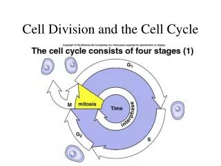
Cell Cycle 14
*Definition: The cell cycle is a series of events within the cell that prepare the cell for dividing into two daughter cells. *Phases of the Cell Cycle: The cell cycle is divided into 2 major events: 1- Mitosis: A short period of time during which the cell divides its nucleus and cytoplasm, giving rise to two daughter cells. It is the shortest period of the cycle taking about 1 hour. 15
2- Interphase: A long period of time during which the cell increases its size and replicates its genetic material and centrioles. Interphase is subdivided into three phases: i. G1 (gap) Phase = Pre-synthesis Phase: Is the longest period of the cycle. It is of variable duration. During the G1 phase, there is an intense synthesis of RNA and proteins, including proteins that control the cell cycle, and the cell volume, previously reduced to one-half by mitosis, is restored to its normal size. ii. S (synthetic) phase: This stage lasts about 8 hours. During this period, the chromosomes and centrioles are duplicated. The cell now contains twice the normal complement of its DNA. iii. G2 (gap) phase = Post-synthesis Phase: This stage is short, about 4 hours. It precedes mitosis. During this stage: ♦Tubulin is synthesized for assembly into microtubules required for mitosis. 16
8h 4h 1h 17
*Cell Function and the Cell Cycle: • The cell does not perform its specialized function either during mitosis or during the S-phase, but in either the G1 or G2 phases. So, for interphase cells to become highly specialized working cells, they leave the cycle, either temporarily or permanently and commonly in the G1 phase. 18
*Classification of Cells According to Their Ability for Renewal: According to the cell cycle concept, body cells are of three categories: • Category I-Non-renewing populations: • These are cells that leave the cycle permanently in order to become highly specialized. They are termed end cells. Theyare not capable of renewal. Examples: The nerve cells of the brain and cardiac muscle cells. 19
Category II- Continuously renewing populations: • These are similarly highly specialized end cells, but they can compensate for their loss. • They are replaced from less differentiated cells of the same family that have retained their capacity for mitosis. • In this way, the total number of cells remain the same although its individual members change. • The unspecialized less differentiated cells of a cell family that retain their ability to divide are called «stem cells". • Examples: Bone marrow stem cells that give the different kinds of blood cells. 20
Stem cells Multipotentialmyloid “stem” cell 21
Category III- Potentially renewing populations: These are highly specialized cells that can return to the cycle in spite of their advanced specialization, so they constitute an exception to the rule. This occurs under certain circumstances e.g. an accident to restore the normal number of cells forming an organ. Cells of this category stop division once the growth, of the organ to which they belong is completed. The best example of such population is the cells of the fully grown liver. 22