Download
1 / 20
200 likes | 376 Views
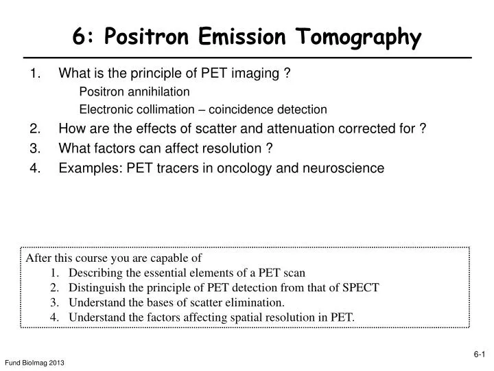
6: Positron Emission Tomography. What is the principle of PET imaging ? Positron annihilation Electronic collimation – coincidence detection How are the effects of scatter and attenuation corrected for ? What factors can affect resolution ?

E N D
6: Positron Emission Tomography • What is the principle of PET imaging ? Positron annihilation Electronic collimation – coincidence detection • How are the effects of scatter and attenuation corrected for ? • What factors can affect resolution ? • Examples: PET tracers in oncology and neuroscience • After this course you are capable of • Describing the essential elements of a PET scan • Distinguish the principle of PET detection from that of SPECT • Understand the bases of scatter elimination. • Understand the factors affecting spatial resolution in PET.
Positron Emission Tomography (PET)Cancer detection (metastasis) in patients Transaxial Coronal PET/CT
Positron Emission Tomography Scanner Detector ring Human scanner PET is similar to SPECT but different (detection principle and instrumentation)
PET of animal modelstransgenic mice, cancer detection micro PET scanner Positioning of the rodent Imaging gene expression (HSV)
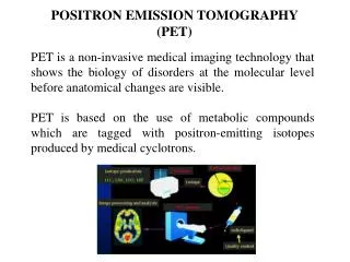
6-1. What is Positron Emission Tomography ? PET • Two issues: • How to determine directionality of x-rays ? • Absorption is undesirable • Positron Emission tomography: measured are x-rays emitted by annihilation of positrons • emitted by exogenous substance (tracer) in body • The principle is as emission tomography, but there is one major difference … (see later) Injection of positron emitter (tracer) - Most widely used tracer for PET 18Fluoro-deoxy-glucose
What does one want to measure with PET ?Annihilation photons Question: Why are two photons are produced? Conservation of linear momentum is not possible with one photon (p=E/c) → 2 photons Energie of photons ? hn=mec2=511keV (1eV = 1.6·10-19 J) - + p→n n Unstable parent nucleus p Annihilation coincidence detection : two events detected at same time → annihilation event along a line (defined by detector) NO need for a collimator NB. Light travels 1m in 3ns : 1[m]/3 108 [m/s]=3ns
What is coincidence detection ?electronic collimation (i.e. w/o physical collimators) Electronic signal What defines simultaneity (coincidence) ? Crystal Photomultiplier Leading edge defines time of detection (sharper, i.e. higher 1st derivative) Bi4Ge3O12 (BGO): t~10ns. Position logic electronics Photomultipliers PET-camera Light guide Scintillating crystal Collimator Elimination of collimator material is a major source of sensitivity increase (why?)
What is really measured with PET ? b b Random coincidences True coincidence a a Scattered coincidence Trues • ) + Rab Tab + Sab (Aab Nab Yab = Scatter What is measured Normalization (Instrument imperfection) Randoms Attenuation
6-2. Why are Random and Scattered Events bad? mimic a true coincidence Random emissions from unrelated nuclear transformations interact simultaneously with the detectors Scatter At least one annihilation photon is (Compton) scattered Erroneous Line of incidence (LOI) assignment to wrong Radon transform Rate of random coincidences : S1 and S2 : count rates on the individual detectors (singles rates) : coincidence time window. Reduce randoms by reducing t (coincidence interval) Does not work for scattered events (why?)
Counts ENERGY WINDOW 700 0 100 200 300 400 500 600 Energy (keV) E = i E f æ ö - q ( 1 cos ) LSO ç ÷ + 1 E ç ÷ i 2 m c è ø e i 2000 425 – 650 How can scattered events be distinguished from true coincidence ? Energy discrimination & background subtraction Measure Ef→ identify severely scattered photons ENERGY WINDOW 511 keV photons Some crystals (BGO) only allow 30% energy discrimination Emission Transmission Scatter BGO Other approaches are needed: 350 – 650 Scatter Corrected Most scattering is by Compton Subtract background (= scatter + randoms) measured in signal void regions → polynomial interpolation
6-3. How is attenuation correction performed ? simpler for PET than SPECT Probability P1: S1=CT(x)*e-m(d-x) P1=S1/CT*(x) CT*(x) Probability P2: S2 = CT*(x)e-mx P2=S2/CT*(x) Attenuation : Probability of detecting the photon pair Compare to geometric average of SPECT (Lesson 5) uncorrected corrected
More attenuation (x4) What are the steps in Attenuation Correction for PET ? PET imaging of the body • Mass attenuation coefficient µ/r in soft tissue = 0.095cm2/g (511keV) • Average path length for the photon pair • longer than for a single photon different lines of response attenuate to varying degrees HVL7cm • Attenuation correction in practice: • Spatially uniform attenuation coefficient assumed • Transmission technique using e.g. Cs source (662keV, why is this good enough ?) Less attenuation single photon emitter Comparison with blank scan i.e. subject removed Correction factor for each Radon transform (µ homogeneous) L
m/r (cm/g) 0.3 0.2 0.1 0 0 100 200 300 400 500 Energy (keV) CT PET Why is PET/CT the industry standard ?PET-Attenuation correction using CT-Data CT + PET = PET/CT CT ~70 keV extrapolation Bone PET 511 keV Soft tissue PET/CT: Two separate scanners in one package scatter & attenuation correction Bone 511 keV CT 6-13 Fund BioImag 2013 Soft tissue
6-4. Why is Resolution never perfect ?Annihilation Range and photon non-collinearity Collinearity: Assumed for Reconstruction Background: At time of annihilation, e-p pair has non-zero kinetic energy Mean free path length (range) conservation of momentum Photon momentum w/zero momentum e-p Range: limits spatial resolution (In air, b+ range ~ several m) pe+pp nucleus + }∆x q ~ 0.25 e- 511 keV 511 keV D (detector distance) D (cm) ∆x (mm) ∆x = 0.5 D tan(0.25) 60 1.3 80 1.7 100 2.2
How does the detector affect PET spatial resolution ? Example: BGO Block Detector Coincidence window: 12 ns Energy resolution: ~ 25% Photomultiplier tube 3 cm • True coincidence count rate RT • RT=2C*TG2 • CT* - tissue activity of a voxel • : the intrinsic detector efficiency (1-e-x) • G : the geometric efficiency (solid angle defined by the detector surface/4). • NB. = 0.9→ 81% of photon pairs emitted towards detectors produce coincidence scintillator Crystal thickness Resolution is best at the center This is a reason for the 3cm thick crystals used for PET detection.
6-5. What are typical PET tracers ?Oncology and neuroscience Oncology 18Fluoroethyl-Tyrosine (FET) Amino acid transport Deoxy-18fluoro-thymidine (FLT) Proliferation 18Fluoromisonidazole (FMISO) Hypoxia Neuroscience 11C-Methionine Amino acid transport and metabolism H215O Blood flow 18Fluoro-Deoxyglucose (FDG) Glucose metabolism Presynaptic dopaminergic function 18FDOPA Blood Flow 15O-Butanol Benzodiazepine-receptor mapping 11C-Flumazenil FDG or F18 fluorodeoxyglucose O15 Water
PET: Neuroscience Normal Alzheimer’s Raw • Pseudo-color display: • red, yellow: high activity • green, blue: low activity
FDG PET Stomach carcinomaEarly detection of response to treatment Response-Prediction 3 M. after CTx Day 14 responder Before CTx non-responder
Whole-body FDG PET Brain Kidney Bladder normal Malignant Lymphoma
X-ray imaging modalities. OverviewCT, SPECT, PET • Measurement of signal integrated along line of incidence (LOI) (Radon transform) • CT: attenuated incident x-ray beam (direction of beam given by source) • SPECT: emitted single photon (need collimation to determine ray direction) • PET: annihilation photon pair (directionality by electronic collimation) Apply correction to measured Radon transform (attenuation, scatter, etc.) Backprojection or central slice theorem: Finally an image!