Download
1 / 17
170 likes | 573 Views
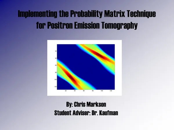
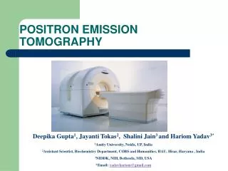
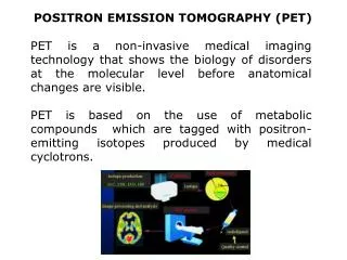
POSITRON EMISSION TOMOGRAPHY (PET). PET is a non-invasive medical imaging technology that shows the biology of disorders at the molecular level before anatomical changes are visible.

E N D
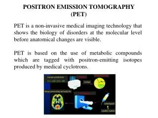
POSITRON EMISSION TOMOGRAPHY (PET) PET is a non-invasive medical imaging technology that shows the biology of disorders at the molecular level before anatomical changes are visible. PET is based on the use of metabolic compounds which are tagged with positron-emitting isotopes produced by medical cyclotrons.
PET/CT The PET/CT system produces directly functional PET and anatomical CT data in one session, without moving the patient and with minimal delay between the reconstruction and the fusion of the two images data sets … CT PET Fused … finally improving the interpretation of PET and CT images
FDG-PET/TC nell'adenocarcinoma del colon retto Ristadiazione per rialzo del CEA Stadiazione pre-chirurgica FDG - PET Risposta al trattamento Caratterizzazione metabolica di lesioni di ndd
FDG-PET/TC nella stadiazione dell'adenok del colon retto T Bassa risoluzione: no informazioni su invasione locale Falsi positivi in caso di: (sospetta lesione sincrona) malattie infiammatorie intestinali polipi Falsi negativi in caso di: tumori < 1cm istotipi mucinosi Doerr RJ et al, Radiology 1998 Kantorova I et al, JNM 2003 Whiteford MH et al, Colon Rectum 2000
FDG-PET/TC nella stadiazione dell'adenok del colon retto N Non è stata provata una maggiore sensibilità della PET rispetto alla TC nell'identificazione dei linfonodi metastatici. Sensibilità PET nei linfonodi addomino-pelvici Doerr RJ et al, Radiology 1998 Kantorova I et al, JNM 2003
FDG-PET/TC nella stadiazione dell'adenok del colon retto Tuttavia tali valori non sono confermati da altri studi: Ridotta sensibiltà della PET nell'identificazione di linfonodi metatastatici adiacenti alla lesione primitiva Furukawa H, Gut, 2006
Donna, 73 anni Pregresso k mammario Nuova diagnosi di Adenocarcinoma del retto (G2)
Donna, 71 anni Adenocarcinoma retto distale In entrambi i pz ln erano stati evidenziati dall''endoecoscopia
FDG-PET/TC nella stadiazione dell'adenok del colon retto M Valutazione resecabilità mts epatiche The changes in the clinical management (%) of colorectal liver metastases are charted on a patient basis for each study and for all studies combined ≥25% Wiering B., Cancer 2005
TC evidenziava due aree ipodense sospette per natura secondaria
FDG-PET/TC nella stadiazione dell'adenok del colon retto M I più recenti studi sembrano attribuire una maggiore sensibilità nella valutazione della malattia extra-epatica addominale e, anche in base all'esprienza del nostro centro, la PET appare più sensibile nell'identificazione della malattia peritoneale
FDG-PET/TC nella stadiazione dell'adenok del colon retto MAddome Torace Capo-collo Polmoni Scheletro Women, seventy years old.
FDG-PET/TC nella stadiazione dell'adenok del colon retto M Indipendentemente dai singoli studi di confronto tra PET e metodiche tradizionali ancora non conclusivi se si aggiunge l'utilizzo della PET a TC, RM e US: 12-27% di cambiamento delle scelte terapeutche dovuto all'aggiunta della PET a TC, RM e US identificando lesioni metastatiche inaspettate o confermando lesioni dubbie Heriot AG, Dis Colon Rectum 2004 Geathart, Ann Surg Oncol 2006 Davey K,Dis Colon Rectum 2008
FDG-PET/TC nella stadiazione dell'adenok del colon retto Evitando l'opzione chirurgica Inducendo ad effettuare chemioterapia neoadiuvante Cambiando il piano di estensione chirurgica Cambiando il campo radioterapico Change in management 10.7% (95% confidence interval [CI] 7.6-14.5%) Vriens D et al, QJNM, 2009
FDG-PET/TC nella stadiazione dell'adenok del colon retto These results suggest that FDG-PET could be used as a complementary exam in case of selected patients showing a major impact in patients with a high metastatic potential, than in case of localized disease. As a result of these considerations, the primary cancer is typically identified by colonoscopy and endo-ultrasonography which allows biopsy and histological confirmation. The extent of disease (primary staging) is accomplished first by CT scan and in some selected patients by FDG-PET. Vriens D et al, QJNM 2009
CONCLUSIONI • FDG-PET non sostituisce le metodiche tradizionali nella stadiazione dl carcinoma del retto • In aggiunta alle metodiche tradizionali impatta sul management di pazienti selezionati con nuova diagnosi • L'indicazione ad eseguire la PET scan in fase di completamento diagnostico è crescente in considerazione dll'utilizzo di tale metodica nella valutazione di risposta al trattamento