Download
1 / 5
50 likes | 152 Views
Explore innovative applications of magnetic carbon-coated cobalt nanoparticles in gene delivery and cell transduction. Learn about site-specific transduction, enhanced gene transfer, and tissue pattern formations.

E N D
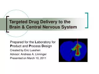
Prepared for the Laboratory for Product and Process Design Created by Eric Lueshen Advisor: Andreas A. Linninger Presented on May 20, 2010 Article Reviews on Magnetic Nanoparticles
Figure 1. Schematic of streptavidin lentiviral vectors conjugated to magnetic carbon-coated cobalt nanoparticles. Figure 2. “U”-pattern formation by magnet-guided transduction of EGFP-transgenic lentiviral nanoparticles. • Streptavidin lentivirals coupled with magnetic carbon-coated cobalt NPs • Magnetically guided to specific sites (Nd-Fe-B magnet) • Transduction into cells (in vitro and in vivo) • Patterns for designed tissue topologies & site-specific transduction
Figure 3. Localization of gene delivery by a magnetic field after 15 min incubation with adenovirus-enhanced PEIbiotin-DNA-SPION formulation. • Superparamagnetic iron oxide NPs coated with PEI + DNA • High transduction efficiency both in vitro and in vivo (GI tract & blood vessels) • Reduced duration of gene delivery to minutes • Host expression of adenoviral vectors in nonpermissive cells • High retroviral concentrations at targeted site
Figure 4 (above). Photo of 5 g air-stable, carbon-coated NPs (lt); TEM image showing 2 to 4 graphene layers coating (rt). Figure 5 (right). Separation of cobalt nanoparticles from a suspension in water at four different times. • One-step, large-scale production of carbon-coated nanomagnets • High air and thermal stabilities • Covalent functionalization with chloro, nitro, and amino linker groups • Excellent magnetic properties (saturation magnetization of 158 A·m2·kg-1)
Figure 6. Ex vivo perfusion of aortas with EGFP-Lentiviral-MNPS showing the localized nanoparticles. Figure 7. Ex vivo perfusion of aortas with EGFP-Lentiviral-MNPs showing clearly visible fluorescence. • Two types of superparamagnetic NPs • Magnetite core coated with positively-charged polyethylenimine • Magnetite core coated with negatively-charged starch with terminal phosphate groups • HIV-derived lentiviral (LV) vectors with eGFP expression promoter • Enhanced gene transfer under nonpermissive conditions • Hydrodynamic flow forces • Low temperatures • Cells transduced with LV/MNP could be magnetically positioned