Download
1 / 25
250 likes | 290 Views
Explore the future of radiology with functional imaging through spectral CT technology with photon counting detectors. Discover the MARS spectral CT project, its goals, team, and capabilities in obtaining novel compositional and functional information about tissues.

E N D
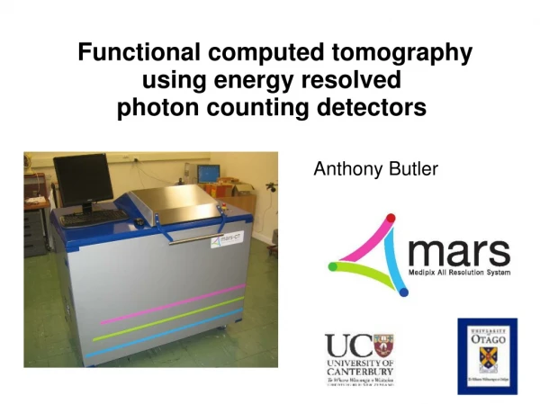
Functional computed tomography using energy resolved photon counting detectors Anthony Butler
Overview Why functional imaging Recent trends in clinical imaging Spectral CT and the MARS project Medical applications Radiopharmaceutical imaging Soft tissue imaging Conclusions
Change in radiology utilisation 1998-2005 => 4.5% /year 2006-2008 => 1.4% /year Bending the Curve: The Recent Marked Slowdown in Growth of Noninvasive Diagnostic Imaging American Journal of Roentgenology, Jan. 2011
Drivers of change 2000-2008 “CT Slice War” • fan beam geometry to cone beam geometry • 2000: acquire a single transverse slice per rotation • 2012: acquire up to 64-500 slices per rotation
Current State Anatomical imaging is now really good Very little benefit in more speed or resolution
Anatomical imaging is now really good Functional imaging is the future What is the tissue? What is its behaviour? Is the treatment working? (not just size, shape, location) What the diagnostician wants to know • Constituents (fat, water, calcium, iron) • Cancer and pathogen labels • Physiological markers • etc
Goals To obtain novel information about tissues Compositional information Functional information To have a route to human imaging
The Team • Technical team • University of Canterbury • Clinical team • University of Otago • International Partners • Incl. CERN, Mayo Clinic, etc • The company • MARS Bioimaging Ltd
Single- , dual-, and spectral CT Single energy CT Xray source B/W Hounsfield Units Grey scale detector Patient
Single- , dual-, and spectral CT Single energy CT Xray source B/W Hounsfield Units Grey scale detector Patient B/W Xray source Dual energy CT B/W Xray source Two grey scale detectors
Single- , dual-, and spectral CT Single energy CT Xray source B/W Hounsfield Units Grey scale detector Patient B/W Xray source Dual energy CT B/W Xray source Two grey scale detectors MARS spectral CT Xray source Medipix Color detectors
Spectral CT is now possible Medipix All Resolution System Energy resolution Spatial resolution Temporal resolution Current single-energy CT provides Spatial resolution Temporal resolution Brightness only (grey scale)
X-ray camera Medipix3 photon processing detector Quantum / counting detector (Film, CR, DR, CT are all integrating detectors) Pixel detector Each pixel has its own electronics Spectral detector Measure energy of photons
Reconstruction tailored to photon counting Photon counting detectors poor in high flux Air Water Ca Sunflower oil Fe Iodine x-ray source Medipix
Reconstruction tailored to photon counting Photon counting detectors poor in high flux Air Water Ca Sunflower oil Fe Iodine High x-ray flux beam x-ray source Medipix High x-ray flux beam
Reconstruction tailored to photon counting Reconstruct only from central detector elements ROI Air Water Ca Sunflower oil Fe Iodine x-ray source Medipix Highx-ray flux
Measure individual materials Iodine: Pulmonary circulation Barium: Lung Calcium: normal bone
Pharmaceuticals identified by spectral information Iodine: Pulmonary circulation Barium: Lung Calcium: normal bone
Functional cartilage imaging Histology and spectral CT to demonstrate GAG content • Low GAG • High hexabrix Cartilage Bone • High GAG • Low hexabrix - Volume rendering - Energy gradient by PCA Funded by NZ Arthritis Foundation
Quantification of fat and water Spectral CT of a mouse 10-35keV “Water-like” “Fat-like” “Calcium-like” Initial work funded by Health Research Council
Atheroma characterization Aim to indentify plaque components Unstable plaques need therapy Next Steps: Ca versus Fe Inflammatory markers Funded by National Heart Foundation
The future: Functional labels • Complex physiological markers can be made • These often have unique spectral response (contain heavy atoms) We can measure the spectral response of nano-particle that target aggregated platelets. Next step: Measure them in mice…
Conclusion Recently radiology improvements have been speed and spatial resolution Functional imaging is the future of radiology Spectral CT is able provide this information Anthony Butler, M Walsh, P Ronaldson, N Scott, R Zainon, S Geiseg, T Janmale, N Cook, A Opie, R Amir, R Doesburg, N de Ruiter,H Yu, J Bennett, G Wang, T Woodfield, N Cook, P Bones, J Mohr, N Anderson, P Butler